Polycystic Disease: Polycystic Liver Disease
Multiple hepatic cysts may occasionally be found as an incidental finding in older hamsters at necropsy. They are considered to be of congenital origin and due to either failure of fusion of the intralobular and interlobular ducts or failure of superfluous bile ducts to disappear.
Raised, cystic areas of variable size, up to 2 cm in diameter, are present on the capsule and within the parenchyma of the liver (Fig. 3.34). True cysts may also be present in other tissues such as epididymis, seminal vesicles, pancreas, and endometrium. In 1 report, over 75% of hamsters studied had cystic lesions at necropsy and many had lesions at multiple sites. Cysts were most common in the liver and epididymis, followed by seminal vesicles and pancreas. The cysts are thin-walled and contain clear, straw-colored fluid. On microscopic examination, there are multiple unilocular and multilocular cystic areas composed of a band of collagenous tissue and lined by flattened to cuboidal epithelial cells (Fig. 3.35). In the adjacent parenchyma of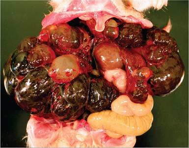
FIG. 3.34. Severe polycystic disease in an aged hamster. There are multiple cystic areas in the liver with compression and disruption of the parenchyma. Source: A. Griffey, Winters, CA. Reproduced with permission from A. Griffey.
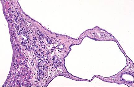
FIG. 3.35. Liver with polycystic disease. The cystic areas are lined by squamous to cuboidal epithelium.
the liver, changes may include pressure atrophy of hepatic cords, hemosiderin deposition, proliferation of bile ducts, and periportal lymphocytic infiltration.