Atrial Thrombosis
Thrombosis, involving the cardiac auricles and atria, is a common occurrence in older hamsters. Either left- or right-sided atria may be involved, but left atrial thrombosis is most common.
Females are usually affected earlier than males, and the syndrome is often associated with amyloidosis. Changes also occur in coagulation and fibrinolytic parameters consistent with consumptive coagulopathy. Atrial thrombosis may be in part due to local blood stasis secondary to cardiac insufficiency. Frequently, there may be concurrent myocardial degeneration and left- or right-sided congestive heart failure.Pathology
Hamsters with this disorder often present with severe dyspnea due to left-sided congestive heart failure resulting from thrombosis of the left auricle and atrium (Fig. 3.32). A moderately firm to friable, pale thrombus may be adherent to the adjacent endocardium. Bilateral ventricular hypertrophy is a common finding. Lungs become congested and edematous. Microscopically,
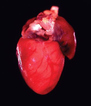
FIG. 3.32. Thrombosis of the left cardiac auricle in an aged hamster. Left auricular thrombosis is associated with left-sided heart failure. The right auricle may also become thrombosed, resulting in right-sided heart failure.
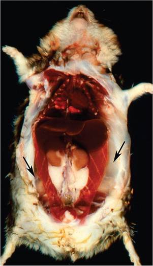
FIG. 3.33. Subcutaneous edema (arrows) in a hamster with rightsided congestive heart failure due to auricular thrombosis.
there may be some degree of organization of the layered thrombus. Focal to diffuse myocardial degeneration, when present, is characterized by nuclear hypertrophy, vacuolation of sarcoplasm, fiber atrophy, and interstitial fibrosis. There may be concurrent focal medial degeneration and calcification of coronary arteries. In the valves, fibrosis and myxomatous change may occur. Thrombosis of the right auricle may also occur, resulting in rightsided heart failure. Affected hamsters have been referred to as “jelly hamsters” because of the marked subcutaneous edema (Fig. 3.33).