Amyloidosis
Amyloidosis frequently occurs in older hamsters, and is a major life-limiting disease in this species. There is a marked variation in the prevalence, depending on the colony under study.
There is an approximately threefold increase in the prevalence of amyloidosis in females compared to males. A “hamster female protein” with functional characteristics similar to amyloid P has been identified in the sera, particularly in female hamsters. Testosterone administration will inhibit the expression of this female protein and reduce the prevalence of amyloidosis in female hamsters. Amyloid deposition may be detected as early as 5 months, but it is much more common in hamsters examined at 15 or more months of age. There may be a drop in serum albumin and a rise in serum globulins. Amyloidosis may be produced experimentally in adult hamsters with regular injections of casein.Pathology
The kidneys are pale with an irregular, granular capsular surface (Fig. 3.30), and affected livers are swollen, with a prominent lobular pattern. On microscopic examination, the liver, kidneys (Fig. 3.31), and adrenal glands are most frequently involved. Other tissues that can be affected include spleen, stomach, testes, and intestine. In the liver, deposition of eosinophilic, homogeneous material is evident around portal triads and within vessel walls, with variable involvement of the sinusoidal regions. Amyloid deposition frequently occurs initially in the glomerular tufts. The early changes may be characterized by the appearance of PAS-positive hyaline-like deposits along the glomerular basement membranes. The early deposits may have the typical amyloid fibrils evident by electron microscopy but may be negative for

FIG. 3.30. Renal amyloidosis in an aged hamster. The kidneys are pale and swollen.
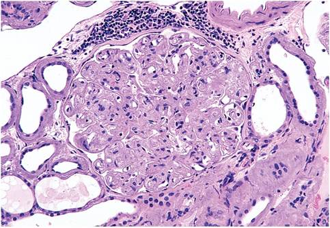
FIG. 3.31. Kidney from a hamster with advanced renal amyloidosis. There is complete obliteration of the glomerular architecture.
amyloid (paramyloid), using the usual histochemical stains. In addition to deposition along glomerular basement membranes, the basement membranes of tubules are also frequently affected. In the adrenal glands, extensive cortical deposition may occur, with distortion of the normal architecture. Atrial thrombosis is relatively common in advanced renal amyloidosis. The loss of antithrombin III in the urine resulting in a hypercoagulable state is considered to be an important predisposing factor.
Diagnosis
The presence of amyloid can be verified using techniques such as Congo red or thioflavin T staining procedures. Deposits may be negative for amyloid using the Alcian blue-PAS staining method. The primary differential diagnosis is hamster glomerulonephropathy.