Polysynaptic reflexes
Tendon reflex
Although the stretch reflex is designed to prevent tearing of skeletal muscle, the tendon reflex, also called the inverse myotatic reflex or reverse myotatic reflex, functions to prevent tearing of tendons (Fig.
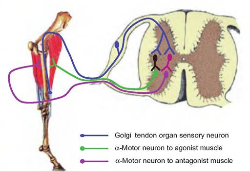
Fig. 10.22. The tendon reflex (inverse myotatic reflex). When the Golgi tendon apparatus within the tendon is stretched, a sensory signal is carried in afferent fibers to the spinal cord where they synapse on interneurons. Inhibitory interneurons synapsing on α-motor neurons going to the muscle where the sensory signal was generated cause relaxation of that muscle. Excitatory interneurons also synapse on α-motor neurons going to antagonist muscles, causing them to contract in order to relieve the stretch on the tendons in the agonist muscle. (Leg was adapted from Riegel and Hakola, 1996.)
10.22). Golgi tendon receptors located within the tendons of the muscle increase their firing rate in response to increased tension in the tendon. When stimulated, afferent signals from the Golgi tendon receptors are transmitted to the spinal cord where these fibers synapse on interneurons. This signal then causes inhibition of contraction of the muscle from which the signal initiated, while causing reciprocal activation of antagonist muscles. The result is relaxation of muscle attached to the overstretched tendon, and contraction of antagonist muscles, an effect opposite that of the stretch reflex.
The Golgi tendon reflex is particularly important during quick activities involving rapid changes between flexion and extension. This reflex is designed to prevent the overstretching of the collagen fibers in tendons.
Withdrawal reflex
The withdrawal reflex, also called the flexor reflex, allows for the immediate withdrawal of a body part in response to painful stimuli (Fig.
10.23). A painful stimulus causes a sensory signal to be transmitted to the spinal cord where it causes excitation of α-motor neurons going to flexors in that region while simultaneously inhibiting the extensors in the same region. This allows for the quick withdrawal of the body part.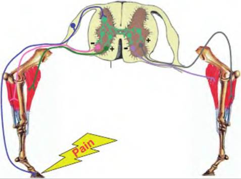
Fig- 10.23. The withdrawal and crossed-extensor reflexes. The withdrawal reflex is initiated as a response to painful stimuli. Stepping on a nail would cause a noxious signal to be transmitted to the central nervous system where it would initiate contraction (+) of the flexor muscles and relaxation (-) of the extensor muscles so that the limb is withdrawn from the painful stimuli. Simultaneously, the crossed-extensor reflex is also initiated, in which the extensors and flexors on the contralateral side are stimulated and relaxed, respectively.
Crossed-extensor reflex
The crossed-extensor reflex is a polysynaptic reflex in which a signal is sent to the contralateral side of the spinal cord to initiate an extensor reflex at the same time the withdrawal, or flexor, reflex is occurring on the ipsilateral side (Fig. 10.23). As an animal withdraws a limb in response to a noxious stimuli, it would fall if it did not simultaneously support itself on the opposite leg. The crossed-extensor reflex immediately allows the animal to support its weight on the contralateral side as it shifts its weight off the ipsilateral side.
Use of reflexes in diagnosis
Reflexes are routinely examined when assessing the nervous system. They provide a diagnostic tool at the site of a spinal cord, brain, spinal nerve, or cranial nerve injury. Muscle tone is tested by passively manipulating a limb. This can provide an indication of whether the animal has hypotonia (less than normal muscle tone) or hypertonia (excessive muscle tone). Disease of the lower motor neurons, those motor neurons with cell bodies in the brain stem or spinal cord, usually causes hypotonia, whereas hypertonia and spasticity are observed with diseases of the upper motor neurons. Upper motor neurons are those neurons with cell bodies in the CNS processing center. When muscles are no longer innervated by the lower motor neurons, they begin to atrophy and lose tone. These neurons either facilitate or inhibit lower motor neurons, which innervate a single motor unit. When an upper motor neuron is diseased, this results in facilitation (i.e., lowering the threshold for propagation of the action potential) of the lower motor neurons, causing hypertonia.
Table 10.3 gives examples of reflexes used in diagnostics. These reflexes are important in determining the site of injury in an animal.