Programmed cell death
Apoptosis is a normal cellular process executed through the activation of conserved cellular signaling pathways that lead to the orderly dismantling of damaged cells marked for death.
Although most fundamental research has characterized differences between apoptosis and necrosis, it is now recognized that cell death can be executed by various pathways leading to the description and study of multiple divergent forms of cell death including ferroptosis, pyroptosis, immunogenic cell death, and necroptosis to name a few (Galluzzi et al., 2018). Stimuli capable of inducing the apoptotic pathway include radiation, hypoxia, nutrient deprivation, and exposure to genotoxic agents (Haupt et al., 2003). Cells undergoing apoptosis adopt characteristic morphologic changes including membrane blebbing, cell shrinkage, and chromatin condensation with orderly nuclear and chromosomal DNA fragmentation. Triggering of apoptosis can be achieved through extrinsic and intrinsic pathways, which converge on a common pathway mediated by executioner caspase enzymes (Figure 3.2) (Zimmermann et al., 2001). 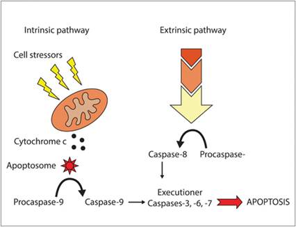
Figure 3.2 Programmed cell death pathways. Initiation of apoptosis is mediated by two distinct yet overlapping pathways with communication between both arms through the generation of truncated Bid. Extrinsic activation is mediated by death receptor clustering, with subsequent proximity activation of extrinsic initiator procaspases (e.g., procaspase-8 activation to caspase-8). Intrinsic activation is stimulated by diverse cellular stressors, including radiation, chemotherapy, hypoxia, and nutrient deprivation, leading to cytochrome c release from the intermembrane space of the mitochondria. Cytosolic cytochrome c binds with Apaf-1 to form a complex called the apoptosome.
Procaspase-9 is recruited to the apoptosome, with consequent proximity activation to initiator caspase-9. Active initiator caspases then cleave and activate executioner caspases-3, -6, and -7, resulting in DNA cleavage and cell death.
The extrinsic arm of the apoptosis pathway is mediated through cell surface receptor clustering mediated by ligand binding and consequent proximity activation of extrinsic initiator procaspases-8 and -10. Cell surface receptors involved in the initiation of the extrinsic cell death pathway include the FAS receptor and tumor necrosis factor-family death receptors (Elmore, 2007; Wang & El-Deiry, 2003; Waring & Mullbacher, 1999). Upon ligand/receptor binding, recruitment of procaspase-8 and -10 molecules to associated cytoplasmic death domains results in the proximity activation of these procaspases to active caspases (Figure 3.2, extrinsic pathway). Fully active extrinsic caspases then proceed to cleave and activate executioner procaspases-3, -6, and -7. Activated executioner caspases cleave multiple substrates, which leads to DNA fragmentation and cell death.
The intrinsic apoptotic pathway, also referred to as the mitochondrial pathway, is initiated following cellular stress or damage to DNA. Mediated predominantly by the DNA damage sensing properties of p53 and consequent transcription of pro-apoptotic proteins such as Bid, Bad, Bax, and Bak, mitochondrial permeability is increased and leads to the leakage of cytochrome c from the mitochondrial intermembrane space. Once cytochrome c is released, it binds with the cytosolic protein Apaf-1, forming a seven spoke-like wheel complex called the apoptosome (Figure 3.2, intrinsic pathway). Once formed, the apoptosome can recruit and bind with intrinsic initiator procaspase-9, with subsequent cleavage of procaspase-9 to caspase-9 through a proximity activation mechanism (Li & Yuan, 2008; Parrish et al., 2013). Like the extrinsic pathway, caspase-9 consequently cleaves and activates executioner procaspases-3, -6, and -7 with the ultimate induction of DNA cleavage and cell death (Yuan & Akey, 2013). Hence, in normal cells when DNA is damaged and unable to be repaired, p53 protein is principally responsible for directing cells into programmed death through the upregulation and expression of proapoptotic proteins such as Bax and caspases, hence serving as a principal safeguard against the survival of cells harboring mutagenic DNA.