Stomach
Normal
The mucosal layer in dogs and cats is comprised of glandular epithelial cells, which typically exfoliate well, particularly with impression smears from biopsy samples (Figure 7.9).
In the fundus of the stomach, two populations of epithelial cells are present: the parietal cells and the chief cells. On cytology, mucosal epithelial cells are usually found in dense aggregates of uniform columnar cells with round nuclei and moderate amounts of basophilic cytoplasm. They often contain apical mucin-containing vacuoles (Figures 7.10a–c). 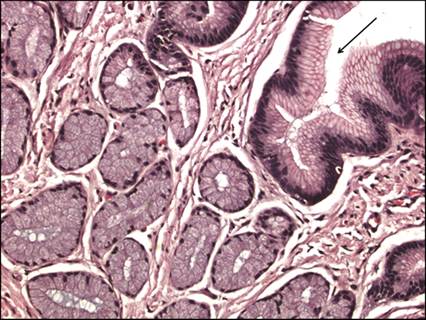
Figure 7.9 Histology section of normal canine gastric epithelium. Note the presence of mucin-containing columnar epithelial cells. Fundic glands, present in cross-section, contain chief cells and parietal cells (H&E, 200? magnification).
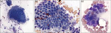
Figures 7.10a–c Normal stomach cytology, canine. (a) Cohesive aggregates of columnar epithelial cells form well-delineated sheets. The cytoplasm is deeply basophilic and often appears vacuolated due to mucin production. (b) Eosinophilic mucin granules are frequently present in aspirates. (c) Small cluster of cells showing normal columnar shape (Wright–Giemsa: a, 200? magnification; b, 500? magnification; and c, 800? magnification).
Hyperplastic lesions
As in other locations, hyperplastic lesions of the stomach are usually presumptively based on diagnostic imaging and the identification of uniform populations of epithelial cells. Mucin production may be increased in some regions and is generally identified as eosinophilic globular material and often found extracellularly. Cytologic samples may appear unremarkable or may be confounded by inflammation; therefore, definitive diagnosis usually requires additional histopathology and investigation for an underlying disorder.
Inflammatory lesions
Infectious gastritis
Helicobacter-associated gastroenteritis has been described in dogs and cats (Neiger & Simpson, 2000; Sapierzyńiski et al., 2006; Jergens et al., 2009). Most research is focused on the incidence of Helicobacter pylori, which is uncommonly identified in the stomach of dogs and cats, although when present it may be associated with more severe clinical signs than infection with other Helicobacter spp. (Neiger Simpson, 2000). The pathogenic role of Helicobacter in dogs and cats is not clear (Rossi et al., 2008; Taillieu et al., 2022). Signs of gastritis can include vomiting, abdominal pain, and inappetence (Sapierzyńiski et al., 2006). Changes to the epithelial mucosa include fibrosis in the lamina propria, degenerative changes in the gastric glands, and hyperplasia of the parietal cells and lymphoid follicles. Cytologically, Helicobacter spp. are identified as spiral-shaped bacterial organisms in gastric samples (Figure 7.11). Evidence of associated inflammation and hyperplasia may be present on impression smears from the gastric mucosa. Organisms are small (approximately 2–4 μm); thus special staining is often necessary to fully identify their presence in tissue sections (Figures 7.12a, b). Speciation requires culture, polymerase chain reaction (PCR) or fluorescence in situ hybridization of biopsy specimens (Jergens et al., 2009). Urea breath and blood tests or serology can be used to diagnose Helicobacter spp. noninvasively but as with other bacterial organisms, presence does not always correlate with signs of disease.
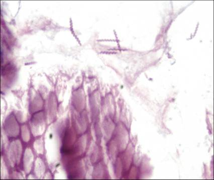
Figure 7.11 Stomach, canine. Tissue section from a gastric biopsy acquired endoscopically. Spiral-shaped bacteria consistent with Helicobacter spp. are present (H&E, 500? magnification).
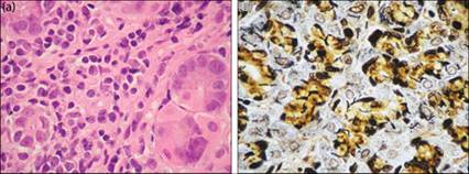
Figures 7.12a,b (a) Severe lymphoplasmacytic inflammation is present (H&E, 1,000? magnification).
(b) Warthin-Starry silver stain highlights spirochetes (Helicobacter spp.) in the lamina propria (1,000? magnification) (7.12a courtesy Al Jergens; 7.12b from Jergens AE, Pressel M, Crandell J et al. (2009). Fluorescence in-situ hybridization confirms clearance of visible Helicobacter spp. associated with gastritis in dogs and cats. J Vet Intern Med 23(1):16–23. Reprinted with permission.)
Gastric neoplasms
Epithelial tumors
Epithelial neoplasms of the stomach are uncommon in companion animals and are nearly always malignant. Rare cases of benign adenomas and hyperplastic polyps have been reported in dogs and cats, with brachycephalic breeds being overrepresented in some canine studies (Gualtieri et al., 1999; Kuan et al., 2009; Kehl et al., 2022). Signalment is typically a young dog with a history of vomiting or regurgitation and weight loss consistent with a pyloric outflow obstruction. Similar to hyperplastic lesions, these usually appear cytologically as normal epithelium, and definitive diagnosis requires histopathology.
Malignant epithelial tumors, carcinomas and adenocarcinomas, uncommonly occur in the stomach of dogs and cats (Gualtieri et al., 1999; Bonfanti et al., 2006; von Babo et al., 2012; Willard, 2012; Munday et al., 2017). In humans, social and environmental factors, such as chronic infection with Helicobacter spp., have been associated with the development of gastric carcinoma. In dogs and cats, the etiology of this neoplasm is not known; however, a hereditary component is reported. Increased incidence of gastric carcinomas is described in specific canine breeds including Chow chows, Belgian shepherd dogs, Bouvier des Flandres, Collies, Standard poodles, and Dutch Tervuren shepherds (Lubbes et al., 2009; Willard, 2012; Seim-Wikse et al., 2013; Koterbay et al., 2020). Similarly, a breed predisposition has been proposed for Persian and Siamese cats (Dennis et al., 2006).
The most successful results for cytologic interpretation appear to be obtained by impression smears from biopsy specimens (Bonfanti et al., 2006); however, FNA is usually successful in carcinomas due to the generally good exfoliation of tumor cells (Figure 7.13). An epithelial origin is usually apparent due to cohesion between cells, which may otherwise bear little resemblance to normal gastric epithelium (Figure 7.10).
Further description of the cytologic features of these tumors can be found below in the intestinal sections. 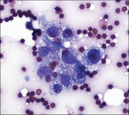
Figure 7.13 Cytology of an FNA of a gastric carcinoma from a cat. A small cohesive cluster of neoplastic epithelial cells displaying criteria for malignancy is present (Wright–Giemsa, 500? magnification).
Stromal/spindle cell tumors
Spindle cell tumors in the stomach are uncommon. Cytologically and histologically, these appear very similar; therefore most were historically, and often erroneously, diagnosed as smooth muscle tumors (leiomyomas and leiomyosarcomas). However, the utilization of immunohistochemical techniques to further classify these lesions has shown that many of these tumors are, in fact, gastrointestinal stromal tumors (GISTs), requiring reclassification of many of those masses (Maas et al., 2007). Several studies have shown distinct localization and prognostic differences between these neoplasms and, therefore, further differentiation by use of specific markers including KIT (tyrosine kinase receptor), smooth muscle actin (SMA), and desmin (a myocellular cytoskeletal element) is often recommended.
In general, spindle cell tumors are usually diagnosed as smooth muscle origin (both leiomyosarcoma and leiomyoma) in the stomach in dogs (Frost et al., 2003; Russell et al., 2007; Gillespie et al., 2011; Hayes et al., 2013). GISTs have uncommonly been identified in the stomach of dogs (Frost et al., 2003; Russell et al., 2007). Rare cases of GIST are reported in the stomach of cats (Morini et al., 2011). Other sarcomas such as osteosarcoma, histiocytic sarcoma, hemangiosarcoma, and undifferentiated sarcoma have rarely been described in the stomachs of dogs and cats. These latter tumors are typically metastases from primary sites and immunohistochemical staining is usually necessary for full characterization of these tumor types (Patnaik, 1990; LaRock Ginn, 1997; Cruz-Arambulo et al., 2004; Bonfanti et al., 2006; Shor et al., 2009).
Identification of stromal/spindle cell tumors in the stomach can be accomplished by cytology, although studies report limited sensitivity with aspiration alone. Alternatively, impression smears have been shown to increase detection from approximately 44% to 100% (Bonfanti, 2006). Therefore, due to a tendency for poor exfoliation of stromal tumors, negative or equivocal results post-aspiration appear to be relatively common. A more detailed description of the cytologic appearance of these tumors can be found below in the intestinal sections.
Round cell tumors
Lymphoma
In cats, lymphoma is the most common neoplasm of the GIT including the stomach (Bonfanti et al., 2006; Willard, 2012). Although lymphoma is also found in the canine GIT, gastric localization of lymphoma in dogs is infrequently described (Bonfanti et al., 2006; Frank et al., 2007).
The cause of gastric lymphoma is unknown; however, recent data suggest an association of alimentary lymphoma with infectious diseases such as Helicobacter spp. (Bridgeford et al., 2008). Although feline leukemia virus (FeLV) infection has been associated with other forms of lymphoma, it has not been consistently linked to the development of the alimentary form in cats (Louwerens et al., 2005; Krick et al., 2008; Lingard et al., 2009; Freiche et al., 2021). A breed predilection in cats may exist as Siamese cats are overrepresented in some studies of feline lymphoma, including primary gastric tumors (Bonfanti et al., 2006; Rissetto et al., 2011). Breed associations occur in dogs as well, although these are most typically noted in intestinal disease as primary gastric lymphoma appears rare in dogs.
FNA and cytology have been shown to be excellent for definitively diagnosing lymphoma in dogs and cats (Bonfanti et al., 2006; Maeda et al., 2017). Primary gastric lymphoma is typically comprised of a homogeneous population of large round lymphoid cells (Figures 7.14 and 7.15). Cells are often fragile and tumor cell rupture is common.
Immunophenotyping of lymphoma has proven useful in predicting behavior in this tumor in most species. Although different morphologic variants of lymphoma are described in both dogs and cats, gastric lymphomas in cats are most likely to be of large B-cell (also called ‘lymphoblastic’) type (Figure 7.14) and typically are associated with aggressive behavior (Vezzali et al., 2010; Moore et al., 2012; Willard, 2012). More discussion on the features and diagnosis of lymphoma is provided in the following section under small intestinal lymphoma. 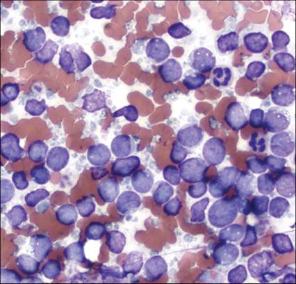
Figure 7.14 Feline, gastric mass. Large lymphocytes typically found in the gastric form of lymphoma. Small, discrete cytoplasmic vacuoles are commonly seen in feline large-cell lymphomas (Wright–Giemsa, 500? magnification).
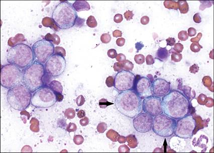
Figure 7.15 Canine, gastric mass. Large, atypical lymphocytes with increased amounts of cytoplasm are found in this patient’s tumor. Small, fine eosinophilic granules (arrows) are identified in several cells characteristic of granular lymphoma, likely of cytotoxic T-cell (CD3+/CD8+) phenotype (Wright–Giemsa, 1,000? magnification).
Mast cell neoplasia
Mast cell tumors may occur in the stomach of cats but are more common in the intestines in this species. In dogs, GI mast cell neoplasia, including gastric tumors, are only rarely described (Ozaki et al., 2002) and usually associated with multifocal masses. Cytologically, they may appear similar to mast cells in other locations but can be variable in appearance. These are discussed more fully in the following section under small intestinal round cell tumors.