The abdominal cavity
The abdominal cavity lies caudal to the thoracic cavity and contains the abdominal viscera (Fig. 2.10). These include the organs of the digestive system and related
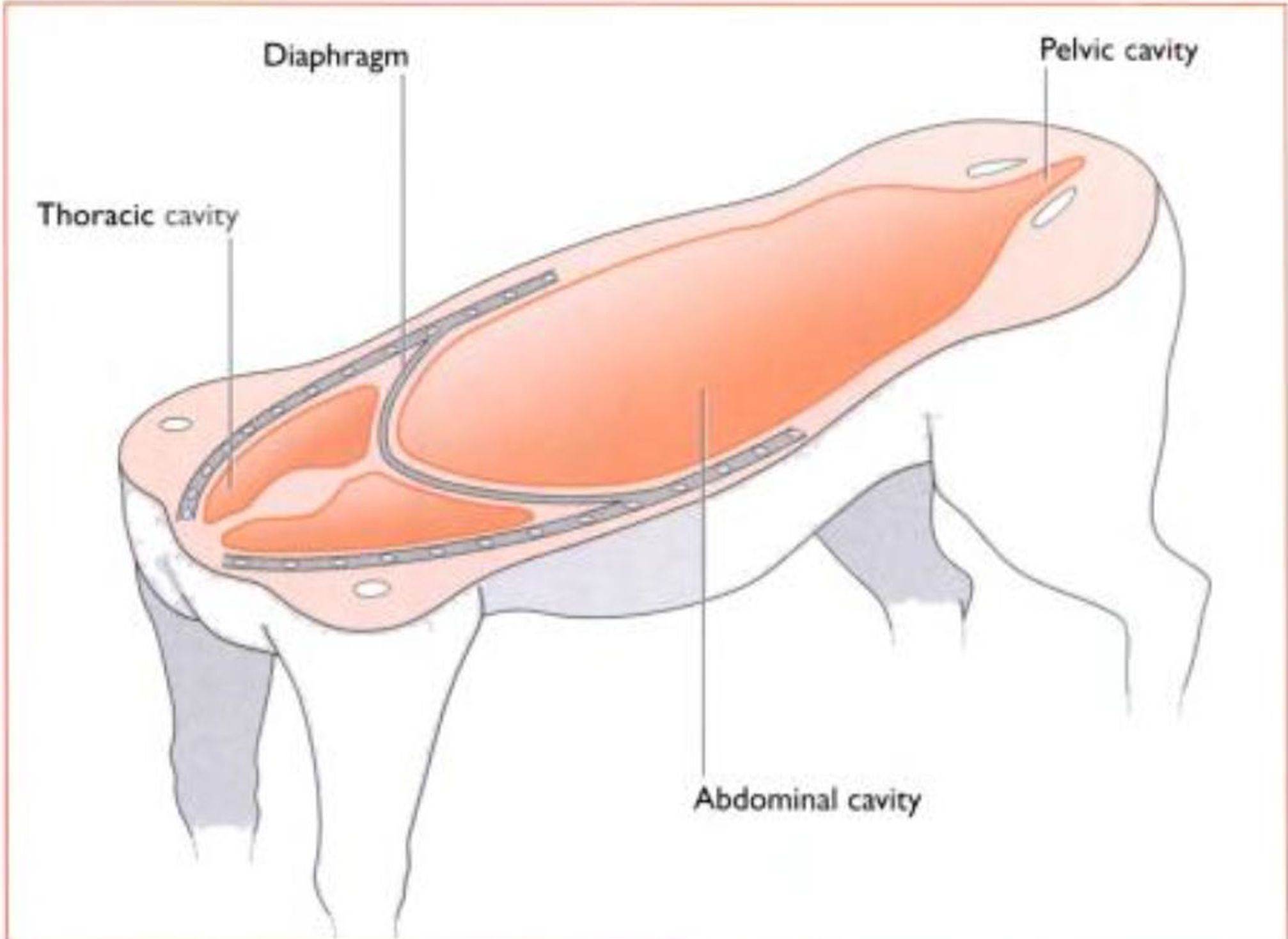
Fig.
2.10 Lo∩g∣iuEhevier Science.)Irregular bones - these have a similar structure to short bones but a less uniform shape: they lie in the midline and are unpaired, e.g. vertebrae.
Some specialised types of bone are:
Sesamoid hones - these are sesame-seed shaped bones that develop within a tendon (and occasionally a ligament) that runs over an underlying bony prominence: they serve to change the angle at which the tendon passes over the bone and thus reduce ‘wear and tear’, e.g. the patella associated with the stifle joint (Fig. 3. 1) Pneumatic hones - these contain air tilled spaces known as sinuses which have the effect of reducing the weight of the bone. e.g. maxillary and frontal bones
Spkiiichiiic hone - this is bone that develops in a soft organ and is unattached to the rest of the skeleton, e.g. the os penis (the bone within the penis of the dog and cal).
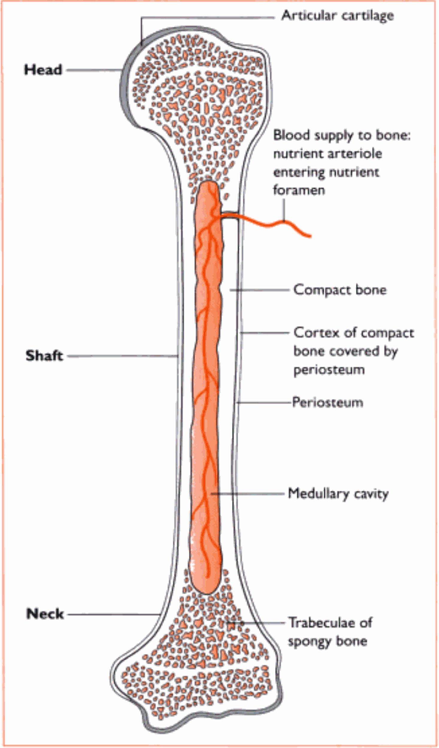
Fig. 3.2 Struaune of a typdrain from the eve into the nose.
Nasal chambers
The most rostral part of Ihe skull carries the nasal chamber. the sides of which are formed bv the maxilla and the roof by Ihe nasal bone. The nasal chamber is divided lengthways into lwo. by a cartilaginous plate called (he nasal septum. Each of the chambers is tilled with delicate scrolls of bone called the nasal turbinates or conchae. These are covered in ciliated mucous epithelium (see Ch. X). At the back of the nasal chamber. forming a boundary between the nasal and cranial cavities, is the ethmoid bone. In the centre of this hone is the cribriform plate - a sieve-like area perforated by numerous foramina through which the olfactory nerves pass from the nasal mucosa to the olfactory bulbs of the brain (sec Ch.
5).The roof of the mouth is called the hard palate, and is formed from three bones on the ventral aspect of the skull: the incisive or premaxilla, part of the maxilla and the palatine. The incisive bone is the most rostral and carries the incisor teeth (Fig 3.5).
Many of the bones of the skull are joined together by fibrous joints called sutures Isee p. O(M)). Sutures are Iirm and immovable joints, but allow* for expansion of the skull in a growing animal.
Mandible
The mandible or lower jaw* is comprised of two halves or dentaries. joined together at the chin by a cartilaginous joint called the mandibular symphysis. Each half is divided into a horizontal part, the body. and a vertical part, the ramus (Fig. 3.6). The body carries the sockets or alveoli for the teeth of the lower jaw. The ramus articulates with the rest of the skull al the temporomandibular joint via a projection called the condylar process. A rounded coronoid process, which projects from the ramus into the temporal fossa, is the point to w hich the temporalis muscle attaches (Fig. 3.6). There is a depression on the lateral surface of the ramus, the masseteric fossa, in wrhich the masseter muscle lies.
Hyoid apparatus
The hyoid apparatus lies in the Intcrmandibular space and consists of a number of tine bones and cartilages joined together in an arrangement that resembles a trapeze (Fig. 3.7). The hyoid apparatus is the means by w hich the larynx and tongue are suspended from the skull. The apparatus articulates with the temporal region of the skull in a cartilaginous joint.
Skull shapes
The shape of the skull varies betwreen species. In the domestic cal the skull is much more rounded or 'apple-shaped* than it is in the dog. and there is little difference between the various cat breeds. In the dog. although the basic anatomy remains the same, the overall appearance differs greatly between the different breeds (Fig. 3.8).
Throe morphological forms of dog skull are recognised:
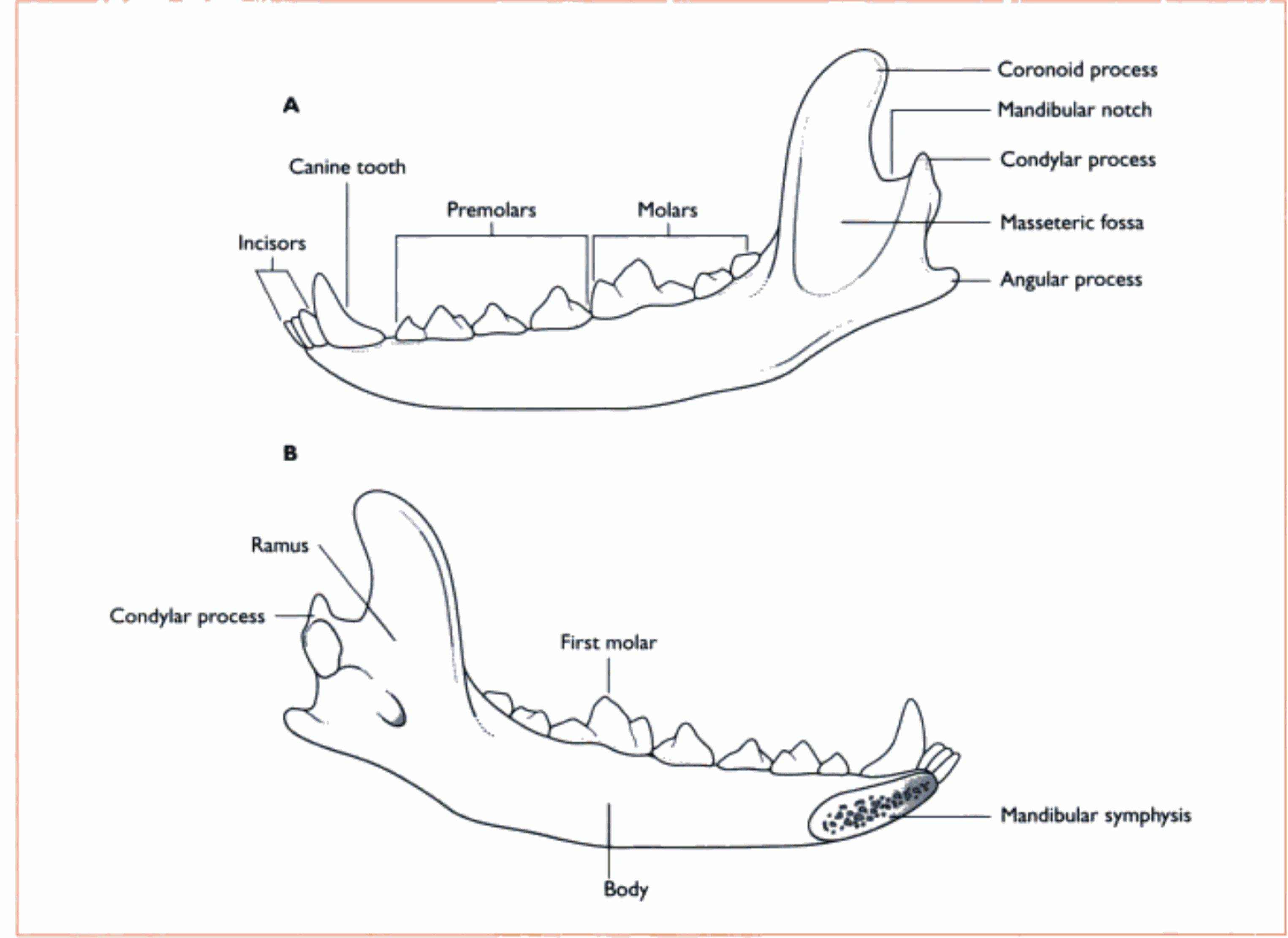
Fig. 3.6 Literal (A) and medal (B) views of the dog mandible.