The eye is (he organ of sight. Photoreceptor cells adapted to respond to the stimulus of light are found in the innermost layer of the eye - the retina.
All mammals have a pair of eyes, each of which lies within a deep bony cavity of the skull known as the orbit. Each eye lies rostral to (he cranial cavity, lateral
| Table 5.2 The autonom∣c nervous system. | ||
| Sympathetic system | Parasympathetic system | |
| Origin of nerve fibres | Vertebrae Tl to L4 or LS | Cranial nerves III,Vll1 IX, X; vertebrae SI,S2 |
| Preganglionic nerve fibres | Short: each nerve leads to a ganglion | Longiganglia lie close to the organ they |
| containing cell bodies and lying close | supply; there is no chain of ganglia | |
| Postganglionic nerve fibres | under the vertebral column; there is a chain of ganglia - the sympathetic chain - one on each side of the vertebral column Long; lead away from the sympathetic | Short; fibres run a short distance from |
| chain and travel towards the organ it | the ganglion to the organ I | |
| Areas supplied | supplies - usually follow the path of blood vessels Viscera in thorax, abdomen and pelvis; | Structures in the head including the eye |
| also supply sweat glands, blood vessels | and salivary glands; vagus (X) supplies the | |
| and piloerector muscles associated with | heart, lungs, stomach, small intestine. | |
| hair follicles. Most ganglia are paired, but | pancreas and large intestine | |
| Transmitter substances: A.Within the system. | three are unpaired: 1. Coeliac - supplies stomach, small intestine, pancreas, large intestine and adrenal medulla2. Cranial (superior) mesenteric - supplies large intestine 3. Caudal (inferior) mesenteric - supplies bladder and genitals Acetyl choline | Acetyl choline |
| i.e. between cell body and dendron B. At the terminal | Noradrenaline | Acetyl choline |
| synapses, i.e. between the axon and the effector organ General effect | Prepares the body for Tear, flight, fight'; | Animal is more relaxed; heart rate is slowed. |
| heart and respiratory rates are increased, | respiratory bronchioles constrict, Gl tract | |
| blood vessels to skeletal muscle are dilated, | activity increases - digestive ∣uices and | |
| blood glucose levels rise, piloerector | salivary secretion increases, peristalsis | |
| muscles to the hairs contract so hackles | increases ' I | |
| are raised, Gl tract activity decreases | ||
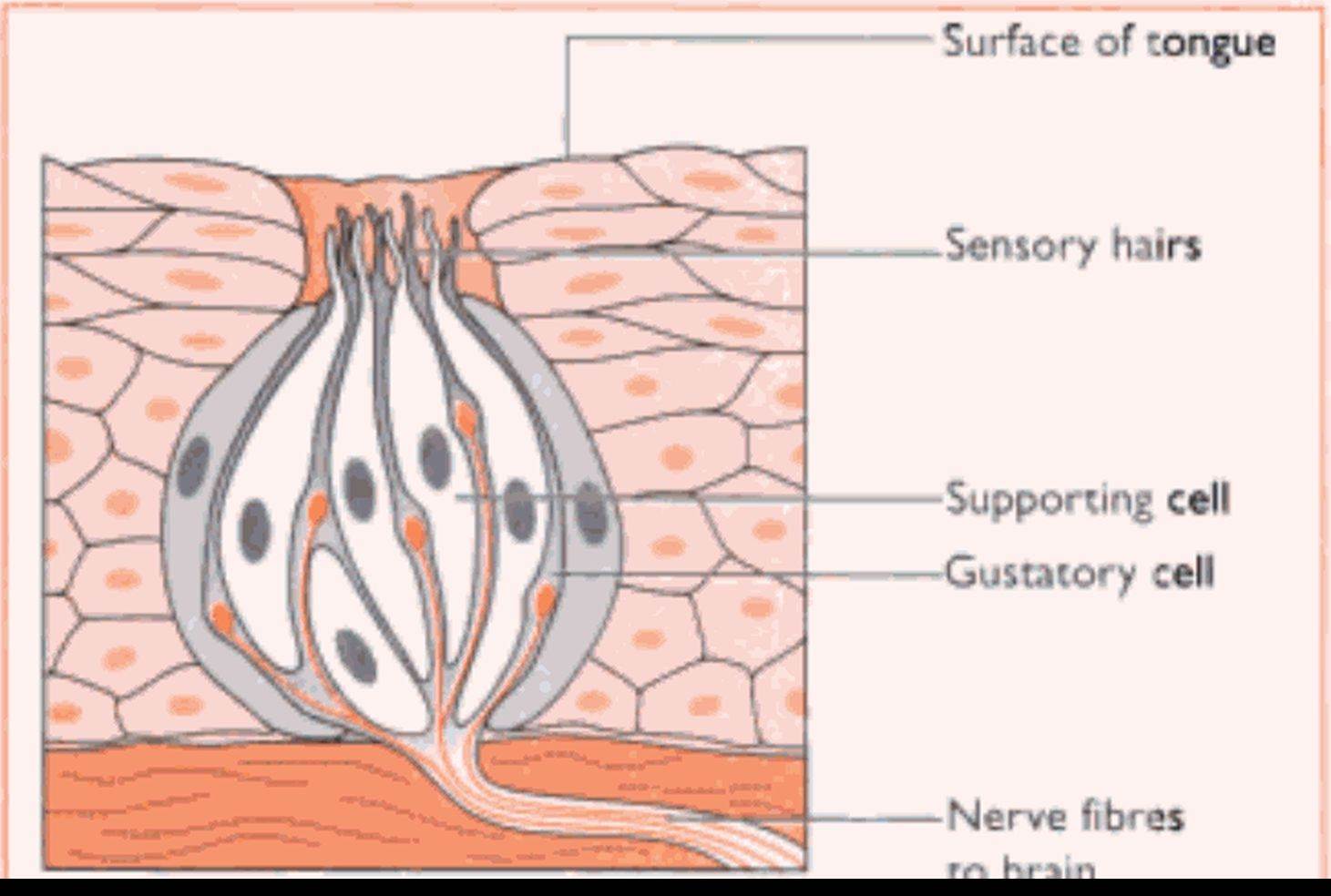
Fig. 5.12 Cross-section through a Uste bud.
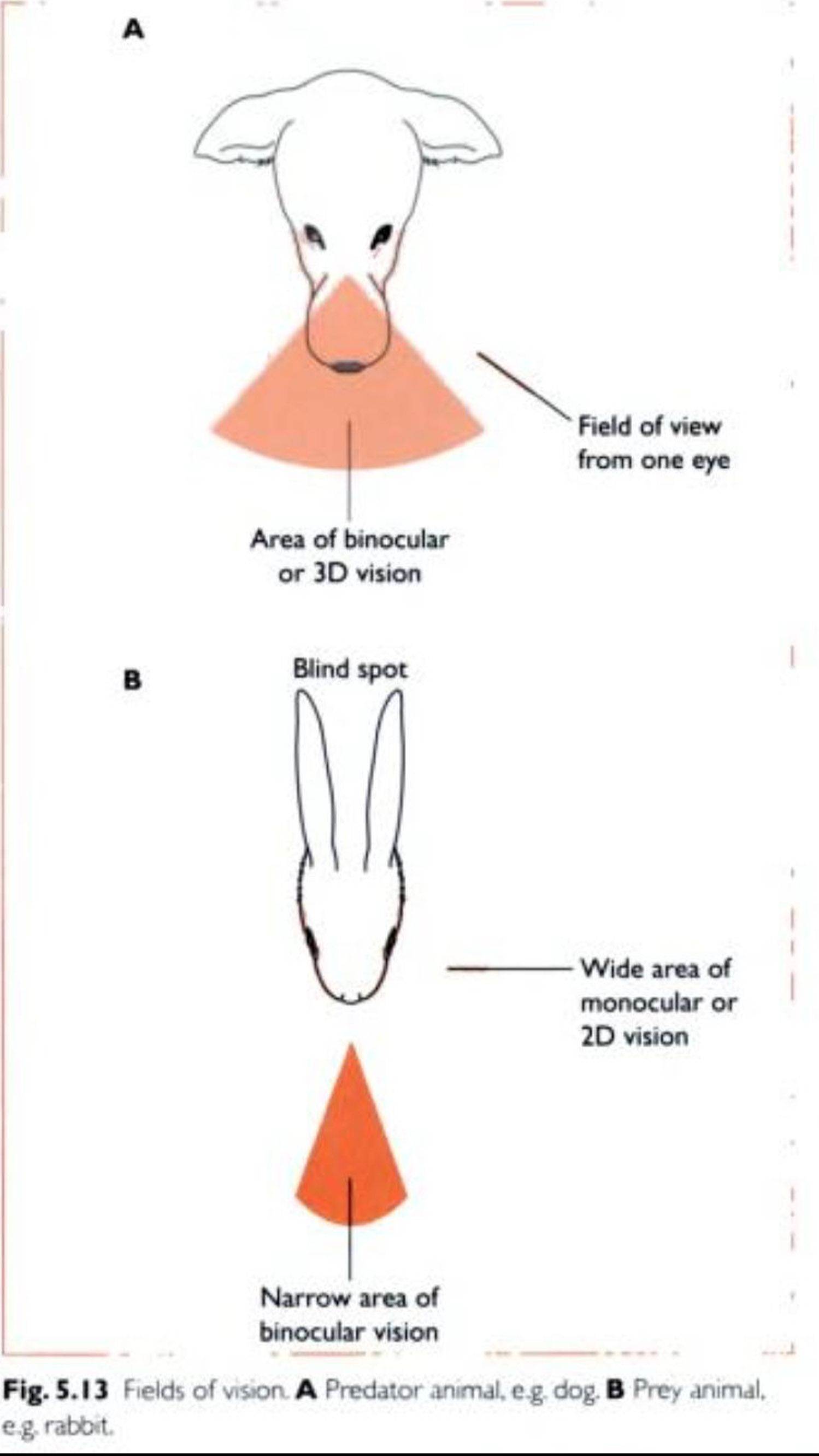
to the nasal cavities and dorsal to the mouth. The dog and the cat are predatory species and their eyes point forwards (Fig. 5.1 5). This provides a wide area of binocular or 31) vision, enabling them to pinpoint the position of their prey accurately. Prey species such as the rabbit or the mouse have prominent eyes set on the sides of the head. These provide a wide area of monocular or 2D vision which enables the animal to see the predator but not to Iix its position - this does not matter. the important factor is that the predator is nearby and that the prey animal runs.
Each eye consists of three main parts: the eyeball, the extrinsic muscles and the eyelids.