The eyeball mg. 5.∣4∣
The eyeball is a globe-shaped Slrucliire made of lhree layers:
1. The s∣ienι
2. 'Γhe uvea
3. The retina.
Sclera
The sclent forms the Iibrous outer covering of the eye.
in conjunction with the cornea.Cornea - covering X of the eyeball. Ihe cornea forms Ihe transparent anterior pari of Iheeye and bulges slightly oulwards from the orbit. Il has a poor blood supply but is well supplied wilh sensory nerve Iibres. The outer surface is covered in a layer of Squamouscpitheliiim. the conjunctiva. The cornea is the Iirsl pari of the eye Io be hit by rays of light and is involved in focusing these on to the retina.
Sc Icni - covering X of the eyeball, the sclera is dull white in colour. It consists of dense Iibrous connective tissue and elastic fibres into which the extrinsic muscles insert. Ils function is to protect the delicate internal structures of the eye and to maintain the eye shape.
The junction between the cornea and sclera is known as the limbus. This is the drainage point for the aqueous humour of the anterior chamber of the eye.
Uvea
The ∣HW∣. a vascular pigmented layer, is Iirmly attached to the sclera at the exit of the optic nerve, but
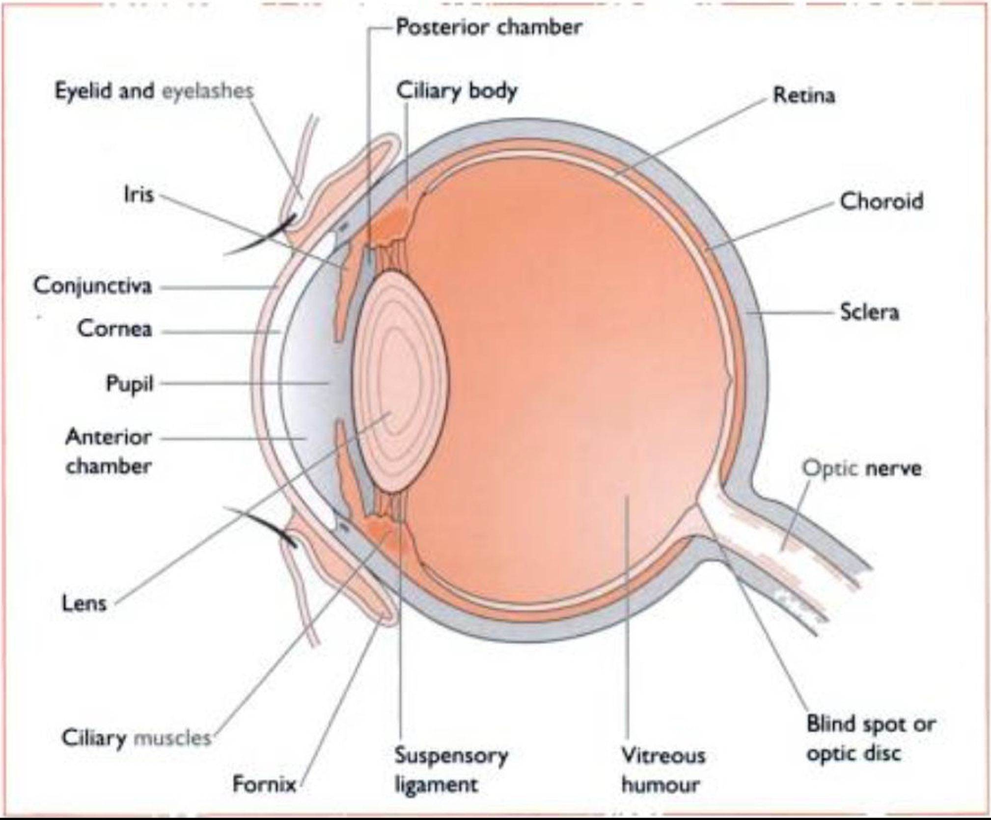
Fig. 5.14 Structure of the can∣nc and Ielme eye
is less well attached in other areas (Fig. 5.14). Il is made up of the following parts:
Choroid - the darkly pigmented lining of the eye which takes up approximately ⅜ of the uvea. It contains the blood vessels supplying all the internal Structuresof Iheeveball.
V
TaprtHin Iiuiduni - a triangular area of yellow-green iridescent light reflecting cells lying dorsal to the point at which the optic nerve leaves the eye (Fig. 5.1 5). Its function is to reflect light back to the Photoreceptorcellsof the retina, making use of low light levels and improving night vision.
The tapetum Iucidum is well developed in carnivores, but present
Fig. 5.15 The retina of the can∙∩e eye. showing the area of the tapetum l∣χ∣dum.
in most Inammalsexccpt mini and the pig. Ciliarij body - a thickened structure projecting towards the centre of the eve. It contains smooth
•r
muscle Iibres - the ι Uiary nιιιs< Ie - which control the thickness and shape of the lens.
Suspensory Iiyiiinent - a continuation of the ciliary body forming a circular support around the perimeter of the lens.
Iris - an anterior continuation of the ciliarv body
∙∙
Conliiining both radial and circular smooth muscle Iibrcs (Fig. 5.14). Its free edge forms the hole in the centre, thepιψil. The dog has a circular pupil while the Ciil has a vertical slit. Pigmcnted cells in the iris give it a characteristic range of colours. The nerve supply to the iris is from the oculomotor nerve (III * and it also receives Sympiithelic and parasympathetic nerve Fibres. The function of the iris is to regulate the amount of light entering the eye.
Retina
The retina is the innermost kιycr of the eye. Light is focused onto the photoreceptor or light sensitive cells of the retina bv the lens and information is transmit- led to the brain via the optic nerve (II). The retina is made of several layers (Fig. 5.16). Light travels through the outer layers before it stimulates the deeper photoreceptor cells.
From the Iavcr closest to the choroid the retina βr
layers are as follows:
Photomeptorivlls- named according Io Iheirshape:
- Rods - sensitive to low light levels but not to colour: provide black and while and night vision
- Cones - sensitive to bright light: provide colour vision.
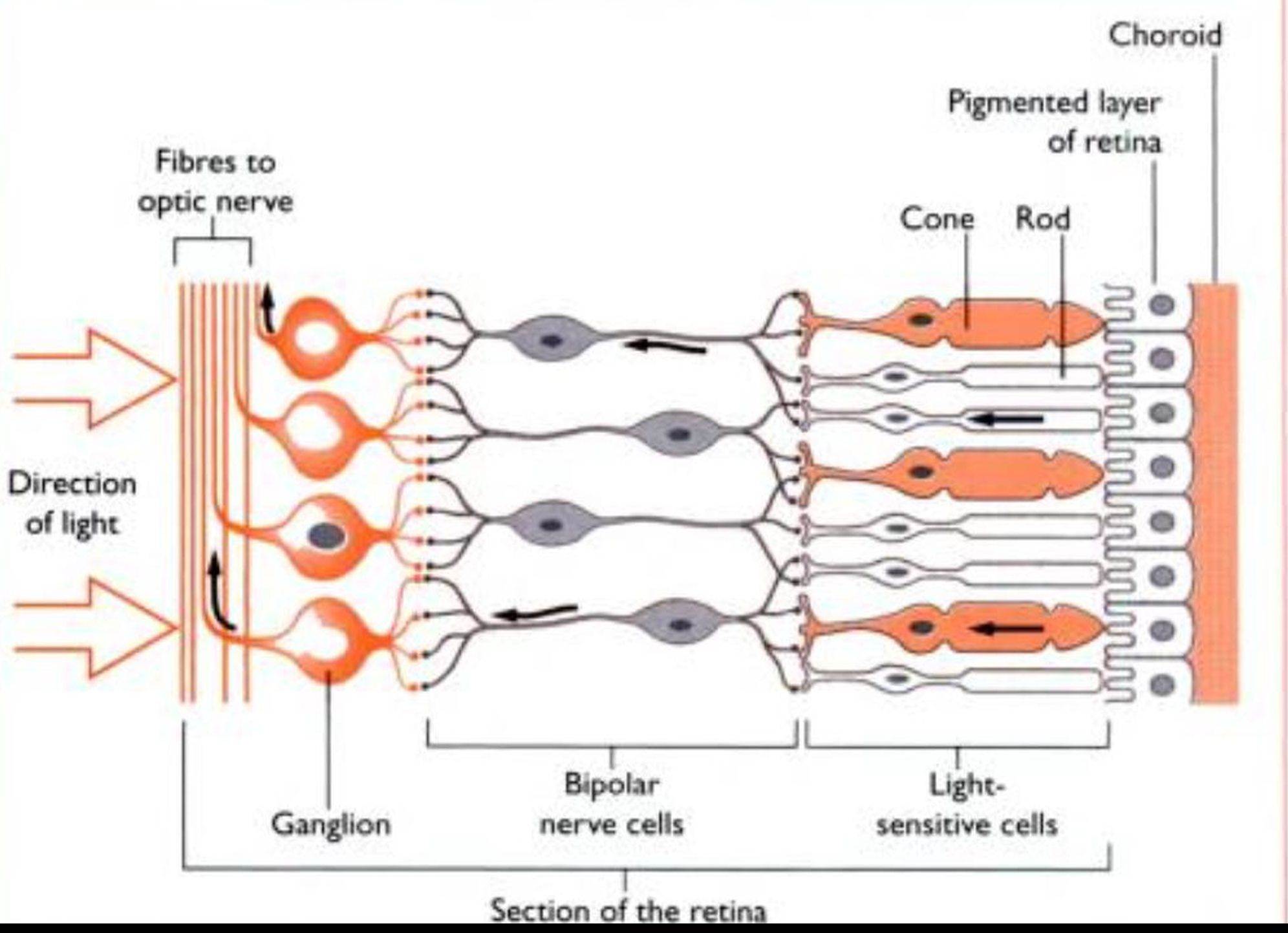
Fig.
5.16 Structure of the retina(cross-section)
The photoreceptor cells of dogs and cuts are made up of 95% rods and 5% cones. This means that they see different degrees of light and shade, but colour vision is poorly developed.
Bipolar neurons - gather information from the rods and cones and transmit it to the next Iaver
W
(iιιnι∣lion cells - may be as many as IxK)6 cells. Their axons travel across the surface of the retina towards the optic disc. where they form the optic nerve till. Al this point there are no rods and cones and for this reason, it is also known as the blind spot (Fig. 5.14).
Within the centre of (he eye is the lens - a transparent biconvex disc surrounded by the suspensory ligament and the ciliary body (Fig. 5.14). The tissue of the lens is crystalline and elastic, and its shape is altered by the contraction of the ciliary muscles around it. This enables (he lens to focus rays of light onto the retina.
The iris divides the eve into (wo chambers:
1. Anterior ( IkihiIht- lies lx∙tween the iris and the cornea, h contains a clear watery Iluid known as ιιt∣ueous humour. which is secreted by Iheciliary body and drains out of the eve via the limbus.
W controlled by the iris in response to the
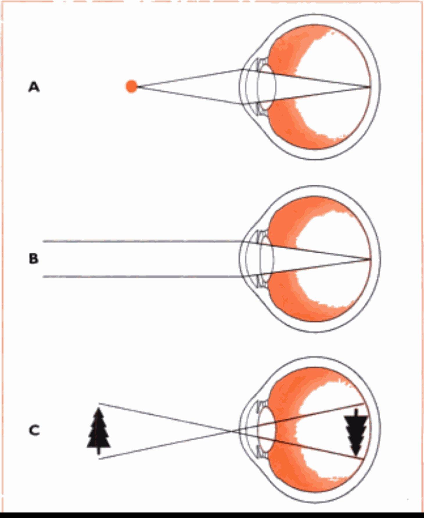
Fig. 5.18 Formation of the √∣ -ual mage. A Light rays From close objects are diverging (spreading out) as they reach the eye.The light ∣s focussed by contraction of the c∣ ⅛ry musc⅛es. which causes the lens to shorten Λ∩d flatten, bending the light onto the reι∣na. B Light rays from distant objects are effectively parallel as they reach the eye.These rays are focus ied by relaxation of the diary muscles, which causes the tens to elongate rd th π CThe ∣πuge OO the retina is upside down.‘Jen it forms; the brain inverts th s, allowmg normal perception of the image the correct way up.
intensity of the light - this is the pupillary reflex. Constriction and dilation of the pupil are controlled bv the autonomic Iiervoussvstem.
? ∙,
k The light rays strike the lens. The curvature of the lens is altered by the ciliary muscles, and the rays converge to a sharp point on the retina - they are said to be fix used.
4. The light rays strike the retina and pass through the layers of cells until they hit the photoreceptor cells. These arc stimulated and send nerve impulses along the nerve fibres which form the optic nerve (111. Information is carried to the visual cortex of the cerebral hemispheres and is interpreted as an image. The image formed on the retina is inverted, but the brain processes the information making use of other sensations, and interprets it as the correct way up.
5. Some of the light is reflected back to the photoreceptor cells by the tapetum lucidum. This makes optimum use of lower light levels.
The ability to aicommotbite. or change the shape of the lens to focus on near and far objects, develops soon after birth. As the lens ages and becomes less flexible.
the time taken for accommodation increases - in man this can be corrected by the use of glasses but animals must Icarn to adapt their behaviour to copc with this ‘old age’ change.