The Integument and Related Structures
Joanna M. Bassert
OUTLINE
| INTRODUCTION, 149 INTEGUMENT, 149 Epidermis, 149 Dermis, 153 Hypodermis or Subcutaneous Layer, 156 Special Features of the Integument, 156 | RELATED STRUCTURES OF THE INTEGUMENT, 159 Hair, 159 Glands of the Skin, 162 Claws and Dewclaws, 164 The Hoof, 164 Horns, 167 |
LEARNING OBJECTIVES
| When you have completed this chapter you will be able to: 1. List the cell types that comprise the epidermis and describe the function of each cell type.2. List the five layers of the epidermis. 3. Describe the process of keratinization. 4. List the structures that comprise the dermis and describe the function of each. 5. List the structures that comprise the hypodermis. | 6. Describe the unique features of the paw pads and planum nasale. 7. Describe the parts of the hair follicle and explain how hair grows. 8. List and describe the three types of hair. 9. Describe the structure and location of sebaceous glands. 10. Differentiate between eccrine and apocrine sweat glands. |
VOCABULARY FUNDAMENTALS
| Anagen phase ahn-eh-jehn faz Anal sac an-ehl sahck Angle ahng-uhl Apocrine sweat gland ahp-o-krihn sweht glahnd Arrector pili muscle ahr-rehck-tor pι-le muhs-uhl Catagen phase kaht-ah-jehn faz Central sulcus sehn-trahl suhlck-uhs Chestnut chehs-nuht Coffin bone kaw-fihn bon Collateral sulcus ko-laht-or-ahl suhlck-uhs Compound follicle kohm-pohwnd fohl-ih-kuhl Corium kohr-e-uhm Coronary band kohr-ah-nor-e bahnd Coronary corium kohr-ah-nor-e kohr-e-uhm Cortex kohr-tehx Cuticle kyoo-tih-kuhl | Defecation dehf-eh-ka-shuhn Dermal papillae dar-mahl pah-pihl-lι Dermis dar-mihs Dewclaw dyoo-klahw Digital cushion dihj-ih-tahl kuhsh-uhn Distal phalanx bone dihs-tahl fah-lahngks bon Eccrine gland ehk-rihn glahnd Epidermal orifice ehp-ih-dar-mahl ohr-uh-fihs Epidermis ehp-ih-dar-mihs Ergot ar-goht Frog frawg Frog corium frawg kohr-e-uhm Hair bulb heor buhlb Hair follicle heor fohl-ih-kuhl Heel hel Hoof hoof |
Hoof wall hoof wahl
Horn tube Oohm toob
Horn hohrn
Hypodermis (subcutaneous layer) hi-po-dar-mihs (suhO-kyoo-ta-ne-uhs la-ar)
Hypophysis hi-pohf-uh-sihs
Implantation angle ihm-plahn-ta-shuhn ahng-uhl
Infraoribital pouch ihn-frah-ohr-Oih-tahl pouch
Inguinal pouch ihn-gwih-nahl pouch
Integument ihn-tehg-u-mehnt
Integumentary system ihn-tehg-u-mehn-tahr-e sihs-tehm
Interdigital pouch ihn-tar-dij-eh-tahl pouch
Keratin kear-ah-tehn
Keratinization kear-ah-tehn-eh-za-shuhn
Keratinocyte kear-ah-tehn-o-sit
Laminae lahm-eh-ne
Laminar corium lahm-eh-nahr kohr-e-uhm
Laminitis lahm-eh-ni-tihs
Langerhans cell lahng-ar-hahnz sehl
Lanolin lahn-o-lihn
Lateral cartilage laht-ar-ahl kahr-tih-lihj
Matrix ma-trihks
Medulla meh-duhl-ah
Meissner’s corpuscle mιz-narz kohr-puhs-ehl
Melanin mehl-ah-nihn
Melanocytehml- e ahn-o-sit
Melanocyte stimulating hormone (MSH) melιl-ahn-o-sit stihm-u-la-tihng hohr-mon
Melanosomehml- e ahn-o-som
Merkel cell mar-kehl sehl
Merkel disc mar-kehl dihsk
Metacarpal bone meh--ah-kahr-pahl Oon
Navicular bone neh-vihck-u-lahr Oon
Notoedres not-o-ehd-rez
Pacinian corpuscle peh-sihn-e-ahn kohr-puhs-ehl
Papilla pah-pihl-lah
Papillary layer pah-pihl-lear-e la-ar
Perioplic corium pear-e-op-lihck kohr-e-uhm
Phalangeal bone fah-lahn-je-ahl Oon
Pheomelanin fe-o-mehl-ah-nihn
Pigmentation pihg-muhn-ta-shuhn
Planum nasale pla-nehm naz-ahl-e
Planum nasolabiale pla-nehm naz-o-la-Oe-ah-le
Pointoypnt
Polled breed polrd O ed
Primary hair prι-mear-e hear
Pruritus proo-rιt-uhs
Quarter kwahr-tar
Reticular layer reh-tihck-u-lahr la-ar
Root root
Root hair plexus root hear plehck-suhs
Sebum se-Ouhm
Secondary hair sehk-uhn-dahr-e hear
Shaft shahft
Sinus hair sι-nuhs hear
Sole sol
Sole corium sol kohr-e-uhm
Squamous cell carcinoma skwey-muhs sehl kahr-sih-no-mah
Stratum basale stra-tuhm Oa-sa-le
Stratum corneum stra-tuhm kohr-ne-uhm
Stratum germinativum stra-tuhm jar-mihn- ah-ti-vehm
Stratum granulosum stra-tuhm grahn-u-lo- suhm
Stratum lucidum stra-tuhm loo-sihd-uhm
Stratum spinosum stra-tuhm spi-no-suhm
Sweat gland —veht glahnd
Tactile hair tahck-tihl hear
Tail gland ta-uhl glahnd
Tactile elevation tahck-tihl e^h-va-
shuhn
Telogen effluvium te-lo-jehn ih-floo-ve-uhm
Telogen phase te-lo-jehn faz
Toe to
Tylotrich hair tι-lo-trihck hear
Tyrosine melanin tι-ro-sen mehl-ah-nihn
Ungula uhng-yuh-lah
Ungulate uhng-yoo-lat
Velvet skin vehl-veht skihn
Vitamin D vι-ta-mihn D
White line whit lin
Wool-type hair wool tip hear
INTRODUCTION
The integument is one of the largest and most extensive organs in the body.
Composed of all four tissue types, it covers and protects underlying structures and forms a critical barrier between the delicate inner workings of the body and the harsh elements of the external world. Its surface is constantly being rubbed, scratched, attacked by microbes, irritated by external parasites, and subjected to environmental chemicals and ultraviolet radiation. The skin, together with related structures such as horns, hooves, claws, glands and hair, form the integumentary system or common integument. This system involves every inch of the external animal and is contiguous with the mucous membranes that line the mouth, anus, and nostrils. It is frequently injured, but possesses a remarkable ability to regenerate and heal.Although derived from living germinal layers, the outer shell of an animal or person is entirely dead. Remarkably, everything you see, from the hair to the skin, is composed of the remains of dead cells. Once alive, in histologically deeper layers and in earlier stages of development, these cells gave up vital organelles and nuclei to make room for the tough, protective substance called keratin. It is during this process, called keratinization, that the cells expire and in doing so form the vital protective barrier that helps enable an animal's survival.
INTEGUMENT
The integument carries out a plethora of protective and regulatory duties. One of its most important functions is to prevent desiccation and rampant infection. In addition, it assists in the maintenance of normal body temperature and excretes water, salt, and organic wastes. It is an important sensory organ that takes in information from the environment via touch and pressure and conveys this input to regions of the central nervous system. It is also engaged in the synthesis of vitamin D and in the storage of nutrients.
The thickness of skin varies among species and according to its location in the body. The thinnest skin, for example, tends to occur around the eyes and the scrotum, whereas some of the thickest layers can be found in the center of the back, between the shoulder blades, and on the paw pads.
Histologically, skin forms two distinct layers: the epidermis and the underlying dermis, which is also known as the corium. These layers are separated by an epithelial basement membrane. In some species, this membrane is wavy and undulating, creating infoldings and outpouchings. The downward folds of the epidermis interdigitate with the upward projections of the dermis, which are called dermal papillae. These interdigitations help cement the epidermis and the dermis together and are therefore most pronounced in areas where there is a great deal of friction.
The epidermis is composed of keratinized stratified squamous epithelium and forms an outer waterproof shield. The majority of skin, however, is composed of the underlying dermis, which is a tough, leathery layer made of dense fibroelastic connective tissue. Only the dermis contains blood vessels. The epidermis is avascular but is provided with nutrient molecules by interstitial fluid that diffuses up from the underlying dermis.
A third layer, the hypodermis or subcutaneous layer, is found below the dermis and is composed primarily of adipose tissue, which acts as a thermoinsulator and a mechanical shock absorber.
Figure 6-1 offers an overview of the structure of skin, which includes the dermis and epidermis, and of the hypodermis with its accessory structures. Let us examine these layers in greater detail.
EPIDERMIS
CELLS OF THE EPIDERMIS
Several different kinds of cell are found in the epidermis. The principal ones are keratinocytes, melanocytes, Merkel cells, and Langerhans cells. The majority of cells found in the epidermis are keratinocytes, which produce a tough, fibrous, waterproof protein called keratin that gives skin its resiliency and strength. Keratinocytes located along the basement membrane are well nourished by the blood supply of the underlying dermis, therefore these cells can grow and divide. As daughter cells are produced, they push older cells away from the life-sustaining nutrients of the dermis and toward the outer layers of the epidermis.
As the older cells travel from the basal to the superficial layers, they undergo profound changes: They fill with keratohyaline granules, lose their nuclei, cytosol, and organelles, and ultimately become lifeless sheets of keratin. This process is called keratinization, and it enables millions of dead cells to rub off or exfoliate daily at no expense to the health of the animal. Remarkably, an entirely new epidermis forms in humans every 7 to 8 weeks.The pigment found in skin is produced by another type of cell, the melanocyte, which is found in the deepest epidermal layers. The melanocyte is octopus-like and possesses long projections that extend outward to all of the keratinocytes in the basal layer. As its name implies, the melanocyte produces melanin, a dark pigment stored in membrane-bound granules called melanosomes. The melanosomes are transported to the tips of the cellular projections, where they are released into the intracellular space and ultimately absorbed by kera- tinocytes. The keratinocytes use the melanin to protect themselves from exposure to damaging ultraviolet rays.
The Langerhans cell is a macrophage specific to the epidermis. Like other macrophages, it originates in bone marrow and subsequently migrates to the skin, where it
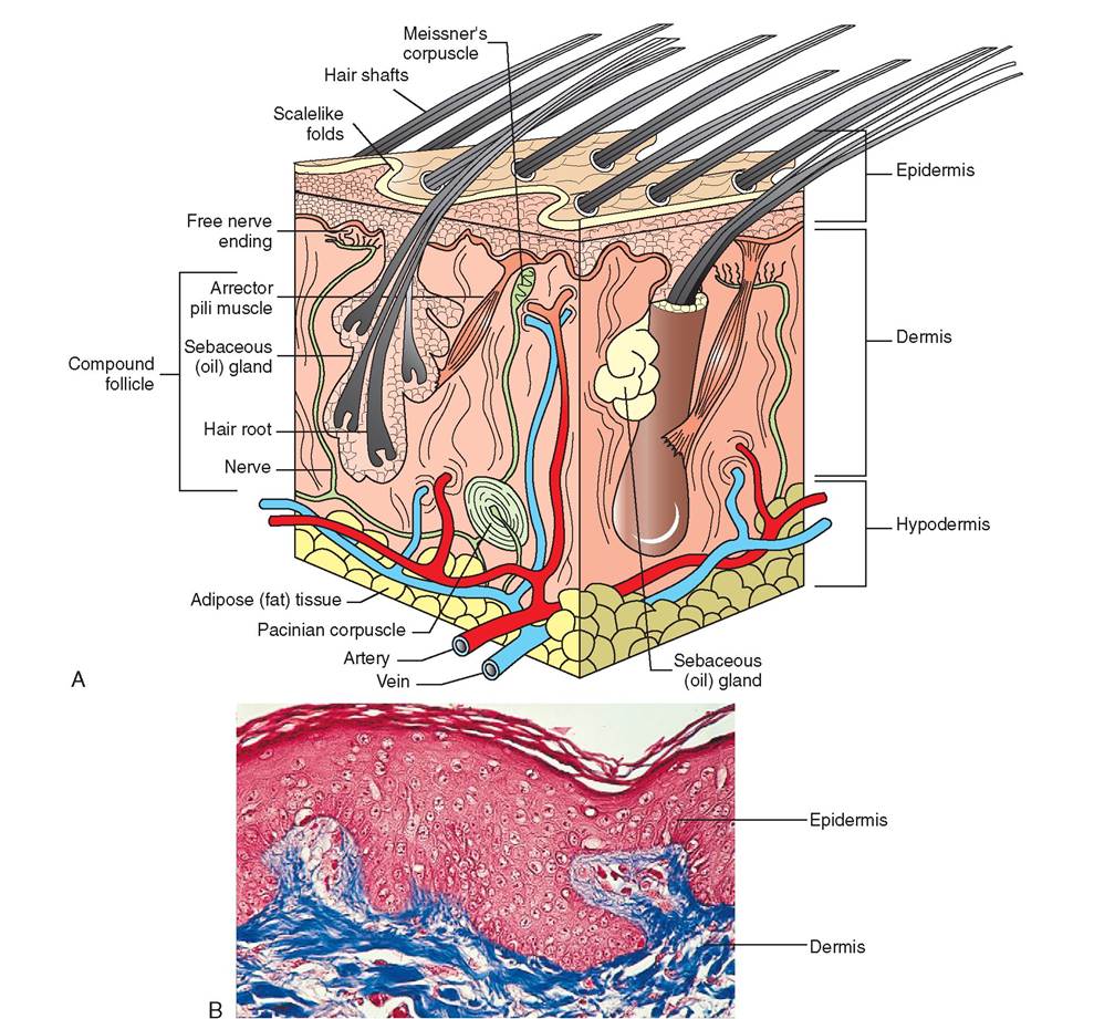
FIGURE 6-1 Canine skin. A, Cubed section of canine skin and underlying subcutaneous tissue, showing many structural details discussed in this chapter. Notice that the epidermis of canine skin includes folds from which compound hairs arise. B, Photomicrograph of skin showing the many cellular layers of the epidermis, the basement membrane, and the underlying dermis. (B, Courtesy Ed Reschke.)
phagocytizes microinvaders and plays an important role in helping to stimulate other aspects of the immune system.
At the epidermal-dermal junction, Merkel cells can be found in small numbers. These are always associated with a sensory nerve ending and are thought to aid in the sensation of touch, taking on a half-dome shape, which perfectly complements the half-dome shape of the sensory nerve ending.
Together, these components form what is called a Merkel disc.LAYERS OF THE EPIDERMIS
Early histologists examined sections of human skin and found five distinct layers in the epidermis (Figure 6-2). These layers were given Latin names and are used today to describe the epidermis in other mammalian species. The deepest layer is the stratum germinativum, but it is also known as the stratum basale or basal layer. For the most part, this layer consists of a single row of keratinocytes, which are firmly attached to the epithelial basement membrane and are actively engaged in cell division. The daughter cells move from the stratum basale to more superficial layers as they mature, to replace epithelial cells that have exfoliated at the skin's surface. Merkel cells, melanocytes, and keratinocytes are found in the basal layer.
The next layer is the stratum spinosum or spiny layer, so named because when the cells of this epidermal layer are
∕ j clinical application
Skin Cancer
With the increasing deterioration of the protective ozone layer that surrounds the earth, people are becoming more aware of the growing risk of skin cancer and the importance of protecting the skin from excessive exposure to the sun. However, we are less likely to consider skin cancer in animals, though cancer of the skin, particularly in certain species and breeds, is very common.
Because cancer is the aberrant growth of cells, skin cancer can stem from any of the cell types found in the epidermis or dermis. As you have learned, many different cell types make up these layers; however, three types of particular importance in cancer are the squamous cells, melanocytes, and basal cells.
Abnormal changes in the genetic programming of melanocytes, for example, can induce a deadly form of skin cancer called malignant melanoma. Malignant melanoma commonly occurs in aged gray horses and initially appears as nodules under the tail base, in the perianal area, and in the scrotum.
Later, these nodules will grow, ulcerate, and spread to multiple internal locations in the horse. Although melanomas may anppear o any area of the body in dogs and cats, they are most malignant in the oral cavity. Among pigs in general the disease iausrter, b malignant melanoma commonly occurs in the Duroc-Jersey breed.Squamous cell carcinoma is another deadly form of skin eccaanuceser, b it spreads rapidly to local lymph nodes and is aggressively invasive locally. It tends to form circular, ulcerated lesions that seem to eat away the surrounding tissue. Squa- emllous c carcinoma commonly appears on the eyeball, nicti- tmatbinragnme, e and surrounding the eyelids of cattle and
horses. It is also seen on the planum nasale, earflaps, and nose ohfitew cats and in the vulvar regions of Merino ewes. It is fone o the most common skin tumors in dogs over the age of 5. Areas of skin that receive prolonged sun exposure are most voulnerable t squamous cell carcinoma.
The basal cell tumor stems from the cells found in the basal lfayer o the epidermis, in hair follicles, and in sebaceous ghleaynds. T do not spread to other areas of the body and teherefore ar considered benign; however, they do recur after removal. Basal cell tumors grow slowly and are found on the head and neck in dogs. They are thought to be one of the most common tumors found in cats but account for only 6% of the neoplasms in dogs.
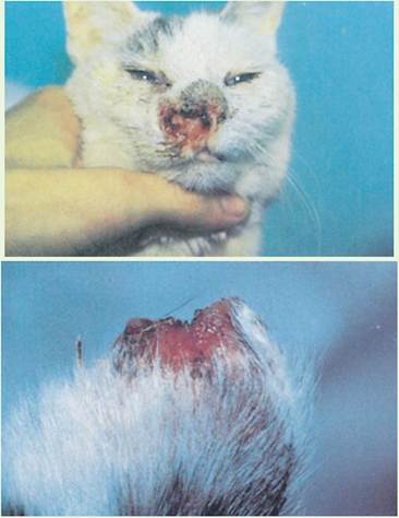
White cats are more vulnerable to developing squamous cell carcinoma. A common location for the tumor to appear is on the nose (top) and on the tips of the ears (bottom). (From Scott DW, Miller WT Jr, Griffin CE: Muller and Kirk's small animal dermatology, ed 6, Philadelphia, 2001, Saunders.)
fixed for histologic examination, they contract into spicu- lated masses that resemble sea urchins. These cells are sometimes called prickle cells-, however, their cellular projections cdcounr ot o naturally, and the cells are normally smooth in
situ. Unlike the stratum basale, the stratum spinosum contains several layers of cells that are held together by desmo- somes. Although cell division is dramatic in the stratum basale, infrequent divisions are seen in the stratum spinosum. Langerhans cells are found in greater abundance in the spinosum layer, where their slender projections form a weblike frame around the keratinocytes.
The stratum granulosum or granular layer is the middle layer of skin. It is composed of two to four layers of flattened, diamond-shaped keratinocytes. The cytoplasm of these cells boegins t fill with keratohyaline and lamellated granules, uwrhnich in t leads to the dramatic degeneration of the nucleus and other organelles. Without these vital parts, the cell quickly dies. The lamellated granules contain water- lpyrcooolifipnidgsg and are transported to the periphery of tehhlele,rec w their contents are discharged into the extracellular space. These glycolipids play an important role in waterproofing the skin and in slowing water loss across the epidermis.
The stratum lucidum or clear layer is only Wnd in very thick skin, so most skin lacks this layer. Microscopically, the stratum lucidum appears as a translucent layer composed of o few rows of flattened, dead cells. In this clear layer and in the outermost epidermal layer, the sticky contents of the keratogranules combine with intracellular tonofilaments to feorarmtink fibrils.
The stratum ^^mιιm or horny layer is the outermost layer and dominates the epidermis. It constitutes up to three fquarters o the total epidermal thickness and is composed of 20 to 30 rows of keratinocytes. Viewed in a sagittal section, the Icnoatmocytes have a paper-thin, almost two-dimensional appearance, yet when viewed from above, they appear hexagonal. Keep in mind that these are really only the remnants eorfaktinocytes, because the actual cell died in the stratum
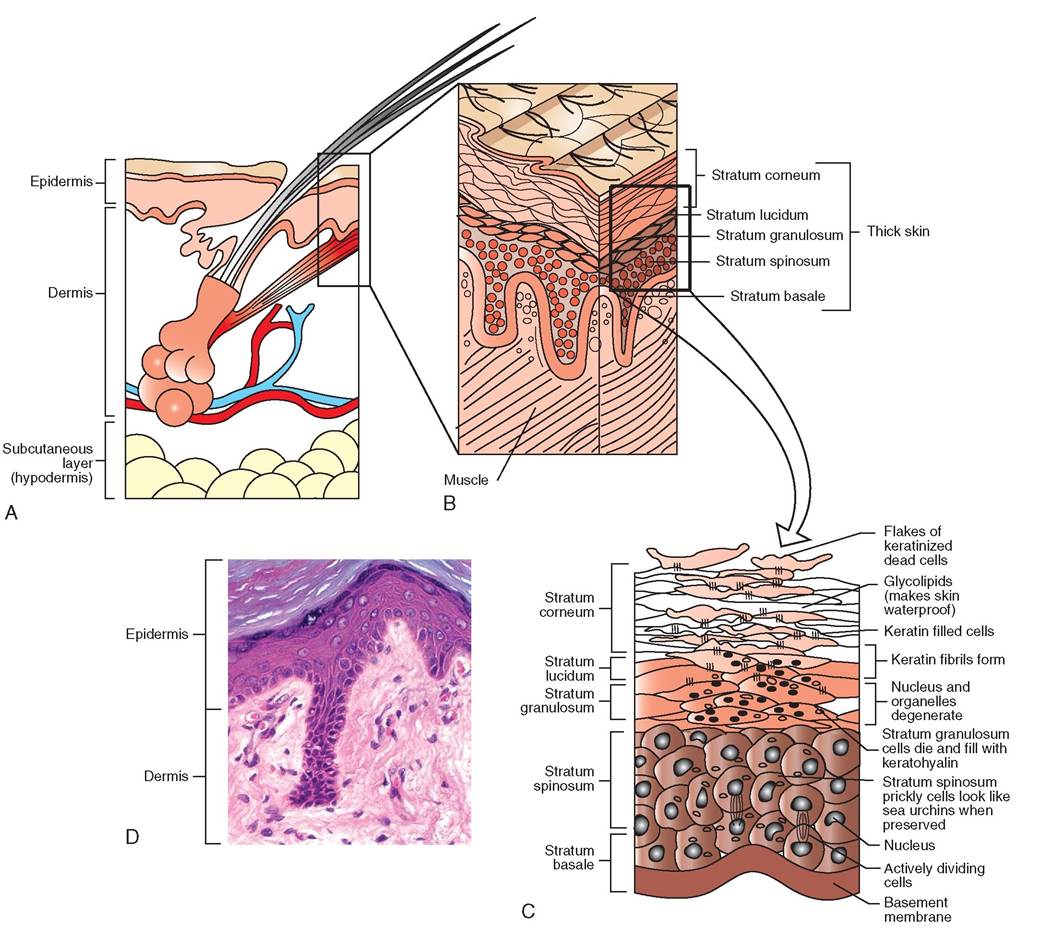
FIGURE 6-2 Layers of epidermis. A, Epidermis is the outermost layer of skin. B, Thick regions of skin are composed of five layers, whereas thinner regions may contain only three. C, Layers of the epidermis. Skin cells actively divide in the stratum basale, where they are supplied with nutrients from blood vessels in the dermis immediately below. As new cells are produced, older ones are pushed into more superficial layers. During this migration, cells lose their organelles, fill with keratin, and die. By the time they arrive at the skin's surface, they have become little more than thin flakes of keratin. D, Light photomicrograph of integument. (D, Courtesy Joanna Bassert.)
granulosum. These remnants are sometimes called horny or cornified cells, but are better known as dandruff.
EPIDERMIS OF HAIRY SKIN
Humans are unusual in their degree of hairlessness, because most mammals are covered with fur. Unlike the epidermis of people and other relatively hairless animals, skin covered with fur usually consists of three epidermal layers, rather than five. These layers are the stratum basale, stratum spinosum, and stratum corneum. The stratum granulosum and stratum lucidum in general are missing. However, a few regions of five-layered epidermis are found in furry mammals, but these are usually seen in regions where the keratinization process has slowed and the skin is very thick.
The surface of hairy skin is covered in scalelike folds. Hair emerges from beneath the scales and is directed away from the opening. In dogs the hair is organized in clusters of three follicles per scale.
Interspersed throughout the surface of the epidermis are knoblike elevations called tactile elevations, or epidermal papillae. Each tactile elevation is usually associated with a tactile hair. These special hairs are called tylotrich hairs, and they are important in the perception of touch (Figure 6-3).
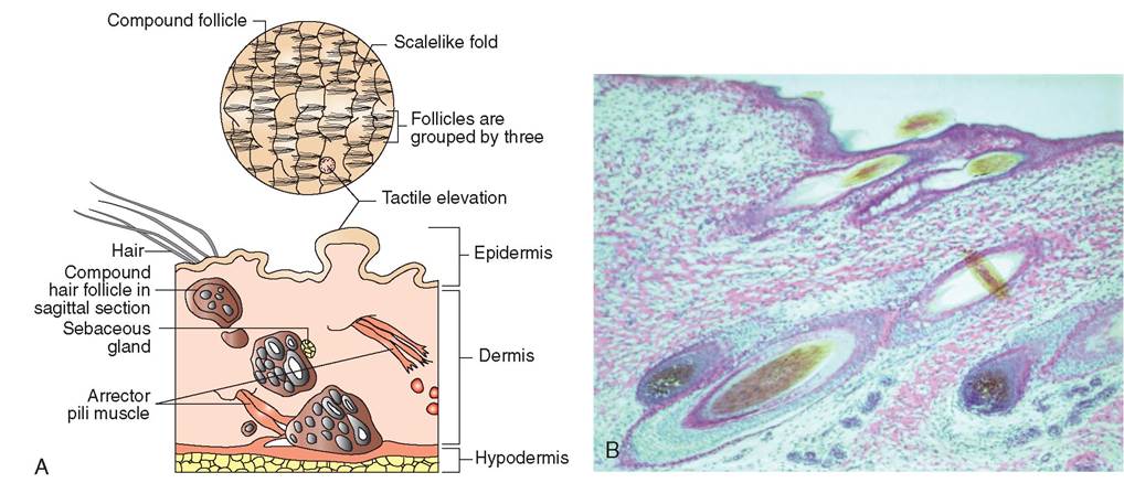
FIGURE 6-3 Tactile elevation and tylotrich hair. A, Compound hairs in dogs are organized into groups of three. Interspersed among these groups are tactile elevations, which are prominent, knoblike extensions of epidermis. They are found in most mammalian species and often are associated with specialized sensory hairs called tylotrich hairs. Hair may be found medial, lateral, cranial, or caudal to a tactile elevation. It is thought that this arrangement enables animals to detect subtle pressure, such as the light touch and movement of insects on their skin. B, Photomicrograph of skin showing hair follicles on the dermis. (Courtesy Joanna Bassert.)
DERMIS
The dermis mates up the greatest portion of the integument and is responsible for most of the structural strength of the skin. Unlike the epidermis, which is primarily cellular, the dermis is highly fibrous. It is composed of dense irregular connective tissue that contains collagen, elastic, and reticular fibers. Hair follicles, nerve endings, glands, smooth muscle, eeslsoeolsd, v and lymphatic channels are all found in the dermis as well, creating a rich and interesting tissue community. Fibroblasts, adipocytes, and macrophages also are present and represent the most commonly found cellular elements in the dermis.
The dermis is a tough layer that binds the superficial epidermis to the underlying tissues. It represents the “hide” of the animal and is the layer often used to make leather. The dermis is composed of two layers: the thin, superficial
CLINICAL APPLICATION
What is Mange Anyway?
Mange is an inflammation of the dermis and epidermis (dermatitis) caused by tiny mites that live on or in the skin. The mites cause irritation, itchmess (pruritus), and hair loss (alopecia). Animals often rub and scratch themselves to the point of causing deep scratches in their skin (excoriations), which ooze serum and blood. The skin thickens (hyperkeratosis) and becomes flaky. Bloody exudates from vigorous scratching harden and form crusts, making the swollen red integument vulnerable to secondary bacterial infection (pyoderma).
Tbe distribution of the red, hairless patches and the way ihnichw the mange spreads depend on the species of the host and the type of mite involved. The type of mange can be identified by scraping the skin with a dull scalpel blade, transferring the scrapings to a microscope slide, and examining the slid: under a microscope. Mites that live in the hair follicles and sebaceous glands are called Demodex. TIiese are long, thin mites with short stubby legs. Demodex are normalIy found in small numbers on many mammals, but in young or immunosuppressed animals the population of mites can go unchecked and increase to abnormal levels, causing visible patches of hair loss. Infestations of Sarcoptes mites are particu- larlu itchy, because they like to burrow into the oozing excoriations of the skin. In contrast to Demodex, Sarcoptes mites huve round bodies. They are drawn initially to areas that are relatively hairless such as the edges of the ears and elbow caps. From these locations, the mange spreads in dogs onto the face, neck, and up and down the legs. Sarcoptic mange is agThe dispersion of the granules is controlled by the release of melanocyte-stimulating hormone (MSH), which in turn is controlled by the intermediate lobe of the hypophysis. The melanosomes are transported to the tips of the cellular projections, where they are released into the intracellular space and ultimately absorbed by keratinocytes. The keratinocytes arrange the melanin on the side of the cell that has the greatest amount of sun exposure. In this way, the pigment acts to protect the keratinocytes from exposure to damaging ultraviolet rays.
PAW PADS
The feet of many animals are padded and quiet. Thick layers of fat and connective tissue form the foundation of the digital pads that bear the weight of the animal. The pad's outer surface is the toughest and thickest skin on the body. It is often pigmented and is composed of all five epidermal layers. Of these five layers, the outermost epidermal layer, the stratum corneum, is thicker than all of the others combined. The insulating fat and tough outer skin form a protective barrier against abrasion and thermal variation, enabling the animal to walk on rough surfaces, hot roads, and cold snow. The surface of the pad feels rough, and an uneven surface is visible with the naked eye. On close inspection, minute conical papillae can be seen covering the entire pad (Figure 6-5). Sometimes the central surface of the pad is worn smooth from walking on rough surfaces such as concrete. In this case, the central papillae are rounded or flattened rather than conical, and the papillae on the periphery of the pad maintain their conical shape and are more grossly evident.
Many species have multiple footpads. These include (1) the carpal pads, which reside on the caudal surfaces of the “wrist,” (2) the metacarpal and metatarsal pads, which are the central weight-bearing pads of the foot, and (3) the digital pads, which protect each of the digits.
In addition to thick adipose layers, the pad is composed of exocrine sweat glands and lamellar corpuscles. Histologically, the ducts from these sweat glands can be seen passing through the dermis to the stratum basale of the epidermis. Their glandular excretion is expelled onto the surface of the pad.
PLANUM NASALE
It is not uncommon for people to judge their pet's health by the animal's nose. An alarmed client, for example, may telephone her veterinarian because her dog's nose is too warm, too wet, too dry, or “just not right.” The top of the nose in cats, pigs, sheep, and dogs is called the planum nasale (Figure 6-6). In the cow and horse, the nose is commonly called the muzzle and is technically referred to as the planum nasolabiale. Like paw pads, the planum nasale represents an unusual form of skin. Although abnormalities in the appearance of the planum nasale can indicate certain illnesses, its wetness or dryness is usually not an indicator of the health of the animal as a whole. Normal animals can have wet, dry, moist, hot, or cold noses. Let's take a look at the planum nasale in greater detail.
On close inspection, the nose of a dog appears to be composed of polygonal plates packed together. Although usually pigmented and appearing as a tough, thick region of integument, the planum nasale in dogs is composed of only three epidermal layers; the stratum lucidum and stratum granulosum are not present. The outermost layer, the stratum corneum, is composed of only four to eight cell layers, which is surprisingly thin considering the exposed location of the nose and its heavy use, particularly in dogs. The epidermal surface is divided by deep surface grooves, which give it the appearance of being composed of multiple plaques. As with other regions of the skin, the dermis and epidermis interdigitate to form an irregular line of attachment that includes dermal papillae. Although often moist from nasal secretions and licking, the planum nasale in dogs contains no glands in
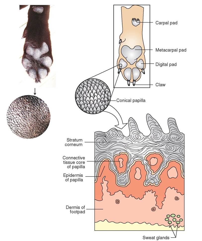
FIGURE 6-5 Paw pads. The pads provide a tough, protective surface on which animals walk. The pad's outer layer is the thickest skin in the body and is composed of thousands of conical papillae. Papillae arise from stratum corneum, the outermost layer of epidermis.
the epidermis or dermis; however, in sheep, pigs, and cattle tubular glands are found.
ERGOTS AND CHESTNUTS
Ergots and chestnuts are dark, horny structures found on the legs of horses, ponies, and other members of the equine family. Chestnuts are usually dark brown and are found on the inside of each leg at the carpus (knee) of the forearm and at the tarsus or hock of the hind leg (Figure 6-7). Ergots are similar but much smaller and are often overlooked, because they are usually buried in the long, caudal hairs of the fetlock. The horse walks only on the third digit, though its ancestors walked on multiple toes much like dogs today. In the course of its evolutionary path, the horse progressively lost digits to become a faster runner. Chestnuts are thought to be vestiges of carpal and tarsal pads of the first digit, and ergots are thought to be vestiges of the second and fourth digits. Visible remnants of a fifth digit do not exist.
TEST YOURSELF 6-2
1. What causes pigmentation of skin?
2. How are paw pads and the planum nasale different from other regions of skin?
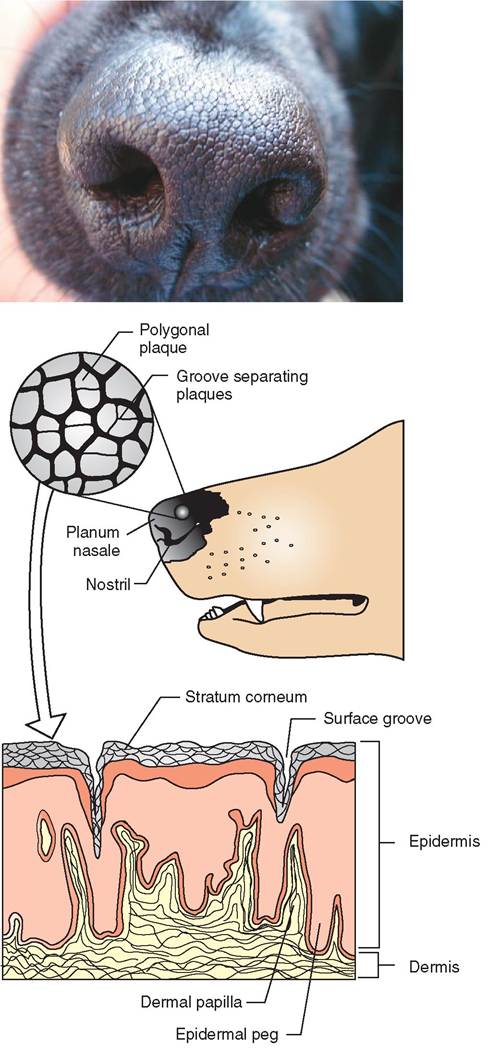
FIGURE 6-6 Planum nasale. The planum nasale is composed of polygonal plaques separated by epidermal grooves. Unlike footpads, the epidermis in the planum nasale is surprisingly thin and contains three layers rather than five.
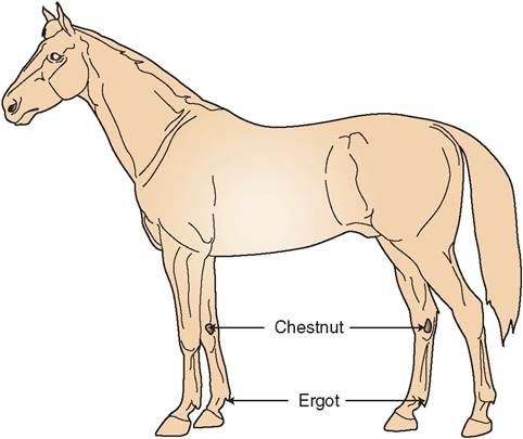
FIGURE 6-7 Location of ergots and chestnuts in horses.
CUTANEOUS POUCHES IN SHEEP
Cutaneous pouches are infoldings of the skin found in sheep. Their three primary locations are in front of the eyes, between the digits above the hooves, and in the groin (Figure 6-8). Respectively, these pouches are technically called the infraorbital, interdigital, and inguinal pouches. Each of these pouches contains fine hairs and numerous sebaceous and oil glands. The glands secrete a fatty yellow substance that dries and sticks to the skin, covering it.
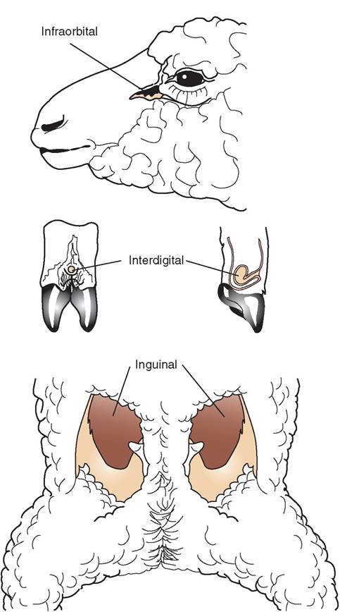
FIGURE 6-8 Locations of cutaneous pouches in sheep.
RELATED STRUCTURES OF THE INTEGUMENT
HAIR
For most animals, hair is essential for survival. By trapping insulating layers of air, hair plays an important role in maintaining body temperature. If dark in color, it can absorb light, which further assists in warming the animal. Coat color may also play a critical role in protecting the animal via camouflage.
In most species of mammal, hair occurs as fur. Marine mammals such as whales, domestic pigs, human beings, and relatively few other species are exceptional in that the hair covering their bodies is sparse and thin. Such species have evolved to survive without fur. For the majority of animals, however, thick fur covers the greater surface of their bodies. Only the hooves, lips, paw pads, horns, nipples, inner folds of genitalia, and nasal regions may be devoid of hair. Animals' coats tend to be thickest on the most exposed regions of the body, such as on the back and sides, whereas the abdomen and inner sides of the proximal limbs are less densely covered.
HAIR STRANDS AND THEIR FOLLICLES
The part of hair that is visible above the skin is called the shaft, and the portion buried within the skin is called the root. Hair is anchored by the hair follicle, which is an invagination of the epidermis that extends from the skin surface to the dermis or, occasionally, to the hypodermis. The deepest part of the hair follicle expands to form a hair bulb. At the base of the bulb is a mound of dermal cells called the papilla, which is covered with rapidly dividing epithelial cells called the matrix (Figure 6-9, A). These cells are nourished by blood flow from vessels in the underlying papilla. Nourishment of the epithelial cells stimulates much cell division and growth. As the cells divide, older cells are pushed upward into the tunnel away from the papilla. These cells become keratinized, and as they lose contact with the nutrition provided by the papilla, they die and become part of the developing hair. In this way hair is constructed from dead epithelial cells.
A web of sensory nerve endings called the root hair plexus envelops the root, making it an important touch receptor when the hair is bent. The wall of the hair follicle is composed of three layers: an internal epithelial root sheath, an outer epithelial root sheath, and a dermal or connective tissue root sheath (see Figure 6-9, B and C).
Animals with fur often have compound follicles in which multiple hair strands emerge from a single epidermal orifice or pore, although each strand has its own follicle and bulb. As many as 15 hairs may be associated with one pore. In compound follicles a single, long primary hair, also known as a guard hair or cover hair, is usually surrounded by shorter secondary hairs, also called satellite hairs. In dogs, three compound follicles are usually grouped together to emerge from the same epidermal fold.
Hair is formed in three concentric layers (see Figure 6-9, D). The innermost layer and central core is called the medulla. It is composed of two to three layers of loosely arranged cells that are separated by spaces filled with liquid or air. The cells themselves contain flexible soft keratin similar to that found in the stratum corneum of the epidermis. Surrounding the medulla is the cortex. Unlike the flexible medulla, the cortex is stiff and rigid because it is composed of hard keratin and is the thickest of the three layers. A single layer of cells arising from the edge of the papilla forms the hair surface, which is called the cuticle. It is also composed of hard keratin. The cells of the cuticle are layered like shingles on a roof, which prevents the hairs from sticking together and forming mats. However in some animals, such as sheep, the edges of the cells in the cuticle are raised, enabling them to “grab onto” the cuticle cells from other hair strands. Because of this, wool threads can be created by twisting and pulling clumps of hair.
GROWTH CYCLES OF HAIR
However unconsciously, we are all aware that hair undergoes a cycle of growing and falling out. When we remove a wad of hair from a clogged sink, when we vacuum up hair left behind by the dog on his favorite chair, and when we can cover the barn floor with a layer of hair from our horse after brushing, we know that it is normal for hair to fall out. Hair is shed to make room for the production of new strands. The volume of shedding is influenced by genetics and by the environment. For example, shedding is heaviest in the spring and fall for animals that live outside. Longhaired animals may shed more than shorthaired animals, and animals kept indoors may shed less than animals kept outdoors. In addition, hormonal changes may influence shedding. For example, many bitches lose a large percentage of their total hair volume at once after whelping. The technical term for this phenomenon is telogen effluvium, but many breeders refer to it as blowing the coat. Whether an animal is undergoing routine shedding or blowing its coat, hair is lost to make room in the follicles for the production of new hair strands. How does this happen?
When a hair is produced, dead keratinized epithelial cells push up and away from the dermal papilla and are organized into the layers that make up the hair shaft and root (Figure 6-10). As more cells are added at the base of the root, the hair lengthens. During this time of growth, the hair is said to be in the anagen phase. As one would expect, the maximum length achieved by the anagen hair is genetically predetermined. In this way, some species and breeds of animals have long coats, whereas others have short coats. When the maximum length of hair is achieved, the hair stops growing, the hair follicle shortens, and the hair is held in a resting phase. This quiescent period is called the telogen phase, which can last from weeks to years depending on the location, type of hair, and species involved. The period of transition between the anagen and telogen phases is called the catagen phase.
HAIR COLOR
Pigment in the cortex and medulla gives hair its color. Genetically programmed melanocytes located at the base of the
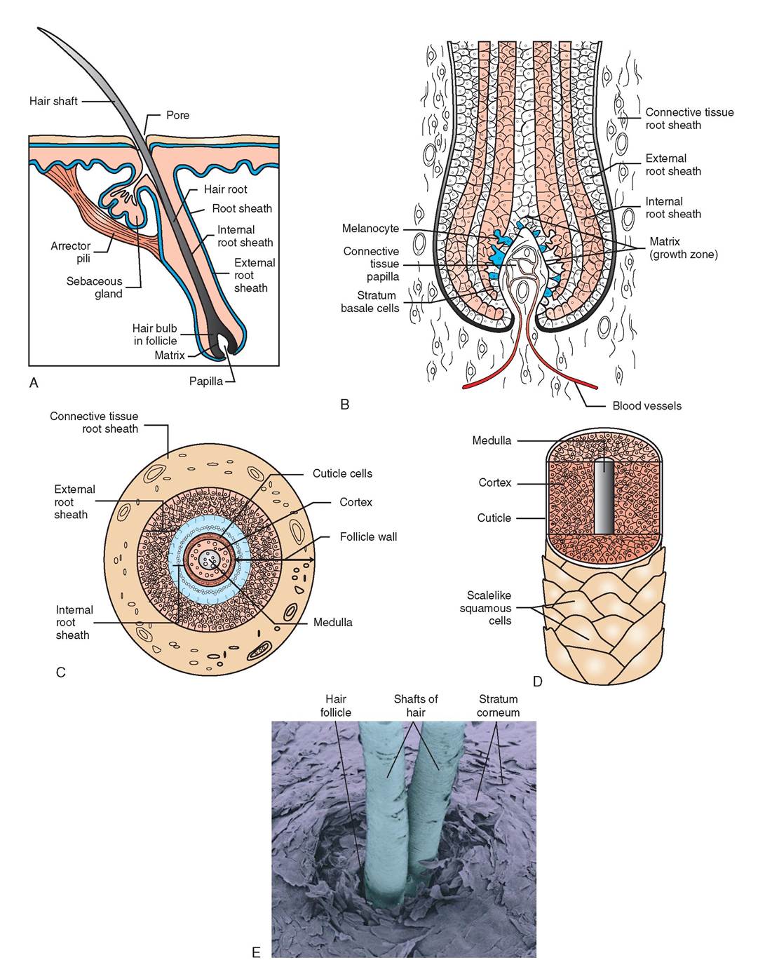
FIGURE 6-9 Hair follicle. A, Structure of a hair follicle. B, The matrix is composed of rapidly dividing epidermal cells that are supplied with important nutrients from the blood vessels in the connective tissue papilla. A strand of hair is formed as daughter epithelial cells mature, fill with keratin, and move away from the papilla and its blood supply. Cells subsequently die and become part of the hair. C, The root sheath is composed of three layers: connective tissue root sheath, external root sheath, and internal root sheath. D, Dead epithelial cells make up the hair. Each strand is organized into three layers: cuticle, cortex, and medulla. E, Scanning electron micrograph of two hair shafts. (Copyright © by David Scharf, 1986, 1993.)
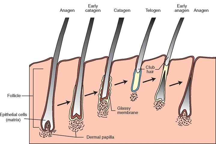
FIGURE 6-10 Growth cycles of hair. The hair growth cycle has three phases. In the anagen phase the follicle is longest. The catagen phase occurs with the appearance of a thick, glassy membrane and a shortening of the hair follicle. Thickening of the basement membrane in the matrix separates epidermal cells from the dermal papilla. In the telogen phase the hair follicle is very short, and the dermal papilla is separated from the bulb. The hair strand is rounded and resembles a club and is therefore called a club hair.
hair follicle produce melanin, which is transferred to the cortical and medullary cells that form the hair strand. Different colors are achieved depending on the quantity and type of melanin incorporated into the hair. Horses, for example, produce only one type of melanin, whereas dogs produce tw. Yellowish and reddish colors in dogs are achieved with pheomelanin, and the brown-black colors are formed by the presence of tyrosine melanin. In horses, all colors are achieved by varying the amount and location of the melanin, not the type. Darker colors are generally iatchhieved w greater quantities of melanin than lighter snhades. I addition, pigmentation may occur uniformly throughout the hair to form a solid color, or it may be concentrated at just the base or just the tip of the strand to form agouti-type coloration.
As animals age, melanin production decreases, and the hair begins to turn gray. White hair is formed when the cortex loses its pigment entirely and the medulla becomes completely filled with air.
TYPES OF HAIR
oAsnsiemssals p a variety of hair types. In general, hair has been categorized into three broad groups: primary or guard hairs, secondary or wool-type hairs, and tactile or sinus hairs. Primary hairs are generally straight or arched and are thicker and longer than secondary hairs. They are the domi- snant hair in a complex hair follicle. As already mentioned, the complex hair follicle in dogs consists of one primary hair surrounded by numerous secondary hairs. Secondary hairs are softer and shorter than primary hairs. They are generally wavy or bristled in dogs and are the predominant hair type in species with wool-type coats. Tactile hairs are usd as probes and feelers. They are well supplied with sensory endings that make them particularly sensitive to the slightest bending or touch. These hairs are commonly known as whiskers and can be found around the mouth and on the muzzle of many species, as well as mixed intermittently throughout tohaet. hair c The tactile hair is also called the sinus hair fbecause o the presence of a large blood sinus, which is located in the connective tissue portion of the follicle.
ARRECTOR PILI MUSCLES
In most animals, hair slopes from the nose to the tail. In seocimese sp or breeds of animal, the hair is more erect than irhnse. othe T degree of erection is called the implantation angle. The summer coats of horses, for example, are short and lie flat against the surface of the skin, therefore the implantation angle in these animals is relatively low. Dog breeds tend to have implantation angles that range from 30 to 40 degrees, although the Chow, Airedale, and Scottish Terrier hwe angles as high as 45 degrees.
Whien frightened or cold, animals can make their hair setyaonnddup b the normal implantation angle. This is due troestehnecep of a small, smooth muscle called the arrector puislcilem,hiwch is attached to each hair follicle and is innervated by the sympathetic nervous system. When the omnutsrcalcets,c it pulls the hair to an erect position.
Perhaps toh have seen a frightened cat “puff up.” This reaction is a defense mechanism designed to make the animal iagpgpeerar b and therefore less vulnerable to potential pred- adntdoirtsio. In, a hair that stands erect can better trap insu-
lyaetrisng la of air than nonerect hair. So animals with erect
∕ j CLINICAL APPLICATION
Allergies: Itchy Business
When cats and dogs develop allergies, they do not usually develop congested sinuses and runny eyes and noses the way oeople do. Instead, dogs and cats develop itchy skin and ears. Lilce people, animals can develop an allergy to just about anything, including human dander. Imagine finding out your pet ls alicrgic to you!
Allergies to inhalant particles such as pollen, dust, and mold spores are common. This type of allergy is called atopy and can cause seasonal itchiness, as in the case of ragweed pollen, or year-round itchiness like that caused by house dust. Atopic dogs tend to rub their faces on the carpet, scratch in the axillae (armpits) with their hind feet, and lick the tops of their p^,s. Food allergies and allergies to ectoparasites, such as fleas, are also very common. Dogs with flea allergies tend to “corncob chew” the base of their tail and the medial sides of their hind legs. Cats rarely chew but exhibit itchiness by excessive licking, grooming, and scratching their face with a aghc.iniadl le F excoriations are evident in the photo on the lxecfet,ssaivned e grooming causes hair loss and redness on the abdomen of a cat in the photo on the right. Notice the lfrimneasl o no skin, which fall into folds when the cat is ckourled t lic its abdomen.
To some ^πt the veterinarian can distinguish between the various types of allergies by the pattern of pruritus or intchiness o the body. Areas that have been scratched or licked excessively will be excoriated, raw, and hairless. In chronic cases the skin may become hyperpigmented and ton ⅛ιk, or areas of white fur may exhibit salivary staining by turning the ehlaloirws.y
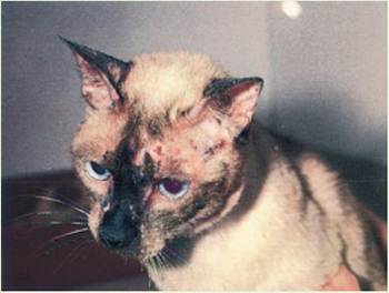
Feline atopy with facial excoriations. (From Scott DW, Miller WT Jr, Griffin CE: Muller and Kirk's small animal dermatology, ed 6, Philadelphia, 2001, Saunders.)
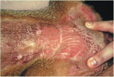
Feline atopy with plaques and alopecia. (From Scott DW, Miller WT Jr, Griffin CE: Muller and Kirk's small animal dermatology, ed 6, Philadelphia, 2001, Saunders.)

TEST YOURSELF 6-3
1. Draw and label the parts of a hair follicle.
2. How doos hairformand grow?
I. What are the fhreo naclar hfhair growth?
4. Why does Fiairturrgtad rah der wMteas animals age?
5. What fectarr shmalate∞htrahair in their ear canals have an increased incidence of otitis externa, which is an infection of the ear canal.
TAIL GLANDS
Most felids (cats) and canids (dogs) possess an oval region at the dorsal base of their tails called the tail gland (Figure 6-12). The tail gland is thought to assist with the recognition and identification of individual animals, and may be grossly recognizable by the presence of coarse, oily hairs. Apocrine and sebaceous glands are especially large in this region. Like sebaceous glands, apocrine glands are sensitive to changes in sex hormone levels, and therefore they become particularly active during puberty and estrus. The tail gland is thought to assist animals in identifying one another.
ANAL SACS
Anal sacs and other related musk glands are famous for their powerful, foul-smelling secretions. Although skunks
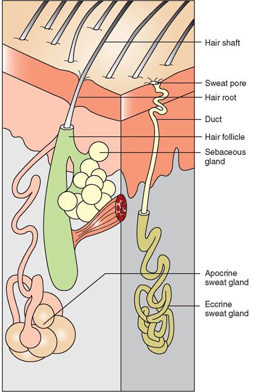
FIGURE 6-11 Glands of the skin. Two types of sweat gland are apocrine glands, with ducts that connect to hair follicles, and eccrine glands, which empty directly onto the skin surface. Sebaceous glands are also depicted.
are shy and not often seen in the wild, it is not uncommon to catch the noxious odor of a skunk's spray from our car as we drive down a suburban or rural road. The odor can linger in the region for days. Cats and dogs have anal sacs similar to musk glands that are located at the 5 and 7 o'clock positions relative to the anus. They are connected to the lateral margin of the anus by a small, single duct. The anal sac is lined with sebaceous and apocrine glands and acts as a reservoir for the secretions that are produced from these glands. When the animal defecates or becomes frightened, some or all of the anal sac contents are expressed, feces become coated with the secretions stored in the anal sac, and the unique smell of the animal is transferred to the environment. Thus defecation serves the purposes of elimination, marking territory, and attracting a mate. Sometimes the small duct of the anal sac clogs and can become infected if left untreated. Animals with irritated or impacted anal sacs often drag their rumps along the ground to help alleviate the discomfort.
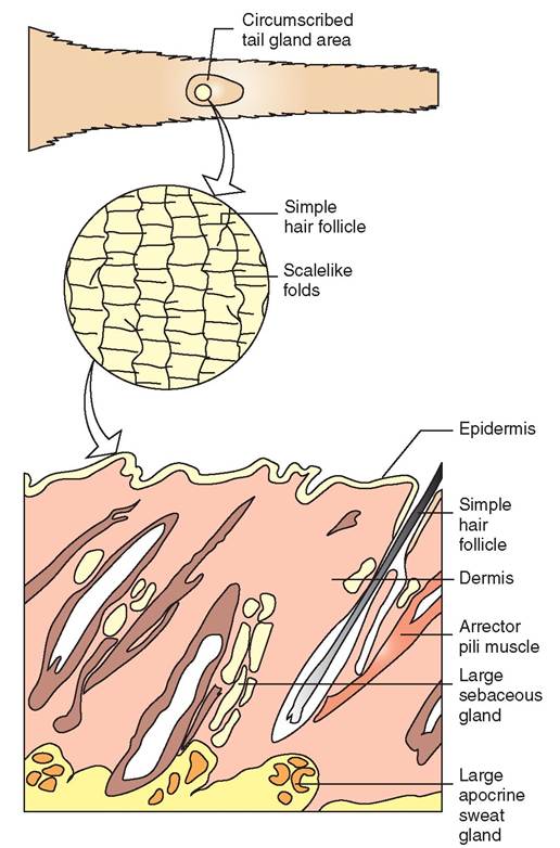
FIGURE 6-12 Tail gland area. The tail gland region in dogs and cats is rich with apocrine and sebaceous glands, which become particularly productive when sex hormone levels are high. Simple, coarse hairs predominate, making the region look grossly different from surrounding areas.
TEST YOURSELF 6-4
1. Name two types of sweat gland. How are they different from one another?
2. Where are anal sacs found and what is their importance to animals?
CLAWS AND DEWCLAWS
Many animals have claws, which are the hard, often pigmented outer coverings of the distal digits. Claws are important for maintaining good traction while running, walking, and climbing and serve as lifesaving tools for defense and for catching prey. In most animals claws are nonretractable, although, with the exception of the cheetah, cats can retract their claws (Figure 6-13). Interestingly, the claws of cats cannot be separated from the distal phalanx bones. A declaw
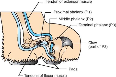
FIGURE 6-13 Cat claw. The claw on a cat is an extension of the third phalanx. Declaw procedures involve complete amputation of this bone.
procedure therefore necessitates amputation of the entire third phalanx. Fortunately, this procedure is usually limited to the front feet.
Dewclaws are the remains of digits that have regressed in the course of evolution. In dogs, the dewclaw is the first digit, but actual bones are only found in the dewclaws of the forelimbs. In cattle, pigs, and sheep, the medial and lateral dewclaws are the second and fifth digits, respectively. Of these three species only pigs have dewclaws that contain bones. Both the metacarpal bones and phalangeal bones are present in the dewclaws of pigs, just as they are in the weightbearing digits.
THE HOOF
We all know what hooves are, but we might not know that the technical name for the hoof is ungula and that hoofed animals are called ungulates. Ruminants have four hooves per foot, and each one covers a digit; however, weight is carried only on two of the four hooves in many ungulates such as sheep, cattle, and goats. The weight-bearing hooves represent the third and fourth digits. Imagine walking only on your middle and ring fingers. In essence, that is what these farm animals are doing. Although their evolutionary ancestors had five toes, the “thumb” or first digit has disappeared, and the “index finger” and “pinky”—the second and fifth digits—have regressed into what we call the dewclaws. These digits are found on the caudal aspect of the foot behind the weight-bearing hooves. As mentioned, a horse, remarkably, walks on only one digit of each foot, that being the third digit, which is equivalent to our middle finger or toe. (See Figure 6-14 for an illustration of the equine foot.)
Both claws and hooves rest on underlying sensitive tissue called the corium. The corium is firmly attached to the periosteum of the third phalanx and is rich with blood vessels that provide nutrient molecules to the developing cells in the inner layers of the hoof. Thus the outer hoof is a modified
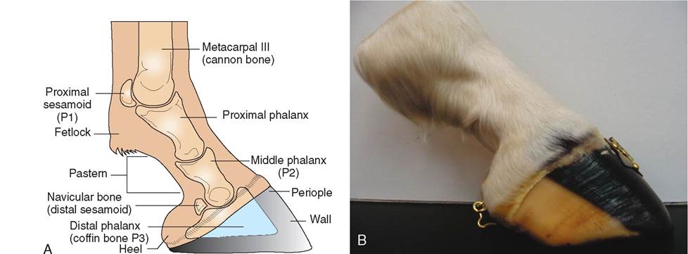
FIGURE 6-14 Bones of the equine foot. A, The skeletal foot of the horse includes the distal part of the second phalanx; the distal sesamoid bone, or navicular bone; and the entire third phalanx, commonly known as the coffin bone. B, Photograph of equine foot.
epithelial layer, and the corium is modified dermis. The corium is well innervated and sensitive to pain, whereas the outer layers of the wall, sole, and frog have no sensation. In addition, the corium is divided into regions based on the portion of the hoof that it produces and/or maintains. There are five types of corium in the equine foot: laminar, periop- lic, coronary, sole, and frog.
1. Laminar corium consists of primary and secondary lamina and is located between the hoof wall and the third phalanx. It provides nutrients to the stratum internum. Laminitis is a serious condition in horses in which the laminar corium becomes inflamed (Figure 6-15).
2. Perioplic corium is located in the perioplic sulcus and supplies nutrients to the overlaying periople.
3. Coronary corium is found in the coronary sulcus and provides nutrients to the stratum externum and stratum medium.
4. Sole corium is located superior to the sole and provides nutrients to the sole.
5. Frog corium is located superior to the frog and provides nutrients to the frog.
The hoof grows from the coronary band downward. Growth of the hoof is continuous, and hooves that are not trimmed can become so long that they curl up like the shoes worn by elves. In wild horses the abrasion caused by running on rough surfaces is an important part of maintaining normal hoof length. Domestic horses, however, rely on a farrier to trim their hooves. Horses are used for work: to carry heavy riders; pull loads; and carry packs, often on hard roads and surfaces. This puts the equine hoof at greater risk for cracking or chipping, which in turn causes lameness and renders the horse unable to work. Long ago, it was discovered that nailing a rigid metal shoe to the plantar and palmar surface of the hoof increased the integrity of the wall and strengthened the foot. A horseshoe prevents excessive expansion of
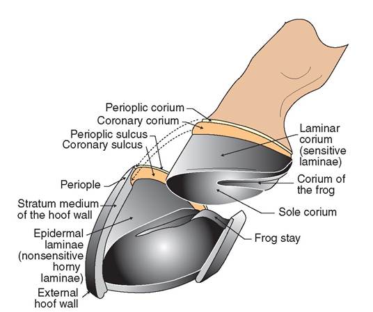
FIGURE 6-1 5 Equine laminae. The hoof is held onto the coffin bone by delicate, interdigitating laminae. When these laminae become inflamed during a painful condition called Iaminitis, the connection between the hoof and the coffin bone is weakened. Consequently, the coffin bone may slip and rotate downward.
the hoof when the animal carries weight. It also improves traction, creates an additional barrier between the hoof and the ground, and allows horses to be kept in working condition with greater regularity.
The skeletal foot of the horse includes the distal part of the second phalanx; the distal sesamoid bone, which is called the navicular bone; and the entire third phalanx, which is commonly known as the coffin bone. The coffin bone is cloaked in a layer of corium, which in turn is covered by the cornified hoof. The hoof and the corium form an
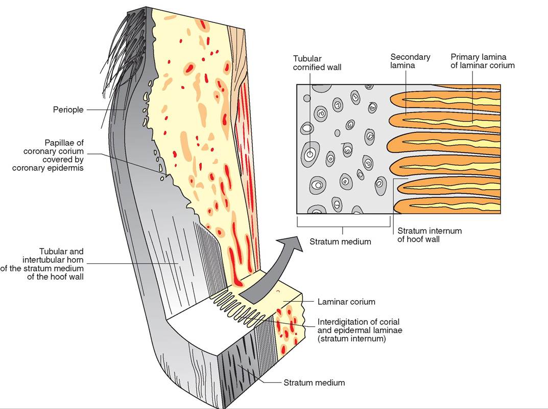
FIGURE 6-16 Longitudinal
cross section of the hoof wall.
elaborate array of interdigitations called laminae. The laminae consist of primary and secondary extensions, which increase the contact area between the corium and the hoof wall. These important interdigitations form the attachment between the hoof and the coffin bone (Figure 6-16).
The equine hoof is generally divided into three parts: the wall, the sole, and the frog. Let's examine each of these parts.
THE WALL
The wall is the convex, external portion of the hoof that is visible from the anterior, lateral, and medial views. It is divided into three regions: the toe, the quarters, and the heels (Figure 6-17). The toe is the front of the foot, and the quarters make up the lateral aspects. The heel is the portion of the wall that tapers downward and wraps around the back of the foot. The hooves of the front feet are angled at about 50 degrees, and those in the back are angled at about 55 degrees. Minute, vertical lines representing horn tubes may be evident running from the coronary band to the ground, and rings or ridges can be seen wrapping around the hoof. Like the rings in tree trunks, these lines or ridges represent periods of growth in the hoof.
THE SOLE
The sole is the plantar or palmar surface of the hoof. It is concave and fills the space bordered by the wall and the bars. The part of the sole that immediately surrounds the bars is called the angle. Like other external portions of the hoof, the outer layers of the sole are avascular and lack innervation. Deeper layers of the corium provide nutrient molecules and contain nervous input. The corium connects the sole to the underside of the coffin bone. A thin strip called the white line is formed at the junction of the sole and the hoof wall.
THE FROG
The insensitive frog is a triangular, horny structure located between the heels on the underside of the hoof. The point or apex of the frog faces the toe, and the base runs across the
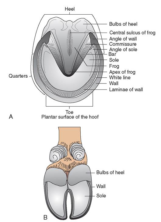
FIGURE 6-17 Anatomy of volar region of equine and bovine hooves. A, Equine hoof. B, Bovine hoof.
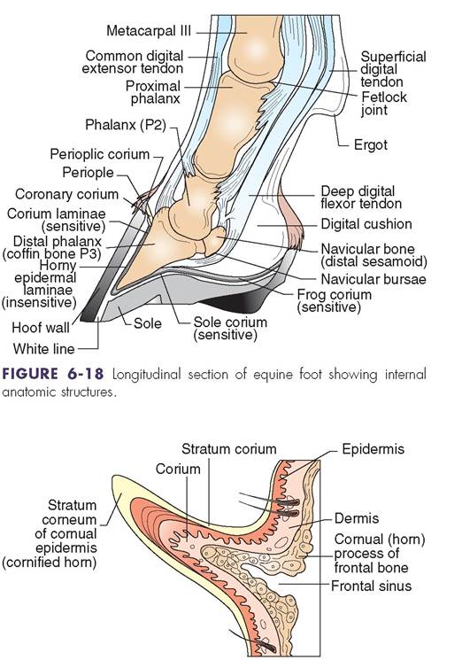
FIGURE 6-19 Cross section through a horn.
caudal aspect of the foot between the heels. The frog is divided by a central depression known as the central sulcus or cleft of the frog. The frog is separated from the bars on the lateral and medial sides by a deep, concave region called the collateral sulcus. A thick pad of fat and fibrous tissue, called the digital cushion, lies beneath the sensitive frog.
Two large bands of cartilage called the lateral cartilages extend proximally from the distal phalanx and form an important structural support for the equine foot. These bands, together with the frog and digital cushion, work as a kind of circulatory pump to assist blood flow through the foot. As the horse bears weight, the frog is compressed against the bars, and the heel of the foot expands (Figure 6-18). In addition, the digital cushion is compressed against the lateral cartilages and the frog, which forces blood out of the corium, away from the foot, and into the digital veins. Shifting weight off of the foot releases the compressive force and enables blood to flow back into the corium via the digital arteries.
HORNS
Like hooves, horns are epidermal in origin and are structurally similar to hair. They emerge from the horn processes of the frontal bones and take on diverse shapes and sizes in ruminating ungulate species such as sheep, goats, cattle, buffalo, and antelope. In adults the horn is generally hollow and communicates directly with the frontal sinus (Figure 6-19). Like the hoof, the horn is a mass of horny keratin. The corium lies at the root of the horn and is bound to the horn process by periosteum. In addition, long, slender papillae of corium interdigitate with one another to form critical attachments that bind the outer horn to the underlying periosteum. The body of the horn is composed of tubules, which are packed close together to form a single mass. Although externally the diameter of the horn is larger at the base and forms a point at the apex, the wall of the horn is actually thinner at the base than at the apex. In fact, the apex of the horn is considerably stronger and denser than the horn base (Figure 6-20).
With the exception of the American Pronghorn, which sheds its horns annually, horns grow continuously throughout the life of the animal and can reach great lengths. Many domestic species of sheep, goats, and cattle are dehorned when young to facilitate their management by the farmer (Figure 6-21). Several different instruments and methods can be used to dehorn an animal, depending on the age and
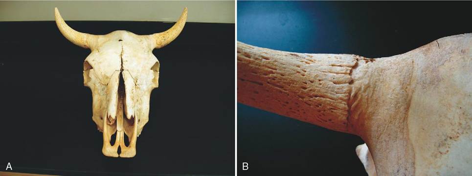
FIGURE 6-20 A, Horns are found on male and female cattle, sheep, and goats and are made of highly keratinized stratum corneum of the epidermis. The medullary cavity of the horn is continuous with the frontal sinus in the skull. Variations in nutrition affect the rate at which horns grow. B, Horns form from the os cornua or horny process, which is an outgrowth of the frontal bone. The horny process is covered with a thick layer of modified dermis called corium, which gives rise to the epidermal cells that make up the horn.
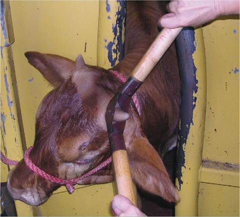
FIGURE 6-21 Dehorning a young calf using a Barnes dehorner. (From McCurnin DM, Bassert JM: Clinical textbook for veterinary technicians, ed 6, St Louis, 2006, Saunders.)
species involved. The standard procedure is to remove the horn Ot Fotii bud and to destroy the corium, usually via cauterization, to prevent further growth. Some species of domestic animals have been bred to be horn free. These breeds are called polled breeds. In nonpolled breeds, horns are found on both males and females. Unlike antlers, they are not sex specific.
on contrast to horns, antlers are found primarily on males, are dermal in origin, and arise as bony protuber- aonmces fr the skull. They grow and are shed annually. Antlers kick a central core and internal blood supply but are nourished externally by a soft, velvetlike tissue. When trhe antle has completed its growth, a dense ring of con- insescuteive t forms at the base of the antler, which restricts blood supply to the outer velvet skin, causing it to die and subsequently be scraped off by the animal. Loss of the lvoewlvset al the antler to harden and become a formidable weapon, a status symbol, and an attractive male secondary sheaar accteristic. With time, the bony connection between
trhe antle and the skull breaks down, the antlers fall off, and new growth begins.
CLINICAL APPLICATION
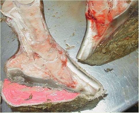
Laminitis: A Painful Health Risk to Horses
Laminitis, or founder as it is commonly called, is an excruciatingly painful disorder that affects the feet—primarily the front feet—of horses and ponies. As its name implies, laminitis is inflammation of the delicate laminae that attach the hoof wall to the underlying coffin bone. As with all inflammation, lami- nitis involves swelling; however, the outer wall of the hoof is rigid and cannot expand to accommodate the swelling of the inner foot, so the laminae become compressed. Blood flow and circulation within the foot are inhibited, and the laminae degenerate. Because the laminae attach the coffin bone to the outer hoof, their degeneration may cause the distal phalanx or coffin bone to pull away from the hoof wall. Under the weight of the animal, the bone may rotate downward and push against the sole of the hoof. With very severe rotation, the distal phalanx can actually perforate the sole, and this will lead to the death of the animal. Chronic laminitis causes abnormal rhowotfhg.
∕ j CLINICAL APPLICATION—cont'd
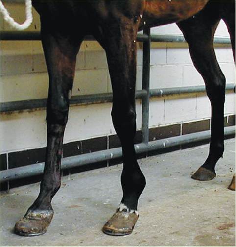
Because Iaminitis is acutely painful, affected animals are often recumbent for extended periods, standing only to urinate, defecate, and access water and food. When standing, horses with laminitis tend to shift their weight away from the front feet to their hind legs to alleviate pressure in the toe. Their gait is slow and hesitant, and their heart rate and respiratory rate may be elevated because of the pain. A mild tap on the toe with hoof testers can elicit a strong pain response from the horse.
In cases of chronic laminitis, external changes to the hoof Vecome evident. Circumferential rings in the outer hoof wall become pronounced, marking previous aberrations in hoof ghreowth. T angle of the hoof is reduced, and the hoof consequently appears flattened. If rotation has occurred, the sole may appear “dropped.” With corrective trimming of the hoof, change in diet, and good management techniques, some of tehrreasentab changes can be corrected.
Predisposing factors for laminitis include the following:
• Engorgement of foods high in carbohydrates
• Any systemic illness or condition that might lead to endotoxemia
• The postoperative period
• Retained placentas in mares •eacAtidovnerse r to drugs
Ponies, in particular, are prone to developing laminitis, particularly if they are permitted to graze on lush pasture or teasrde f die rich in carbohydrates such as corn, molasses, and grains. Tteatment is designed to decrease swelling, relieve pain, eaansde incr circulation in the feet. Prevention by adherence troicta, st low-carbohydrate diet is essential in ponies and horses that are sensitive to carbohydrate levels.
Gross pathologic photograph of sagittal section of both front feet of a horse with bilateral laminitis that has undergone coffin bone rotation. (From McCurnin DM, Bassert JM: Clinical textbook for veterinary technicians, ed 6, St Louis, 2006, Saunders.)
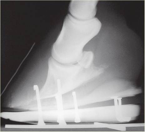
Lateral radiograph of the front foot of a horse with laminitis that shows evidence of coffin bone rotation. (From McCurnin DM, Bassert JM: Clinical textbook for veterinary technicians, ed 6, St Louis, 2006, Saunders.)
Abnormal hoof growth in a horse with chronic laminitis in both front feet. (From McCurnin DM, Bassert JM: Clinical textbook for veterinary technicians, ed 6, St Louis, 2006, Saunders.)