Treponema paraluis-cuniculi Infection:
Rabbit Syphilis
Treponema paraluis-cuniculi (formerly T. cuniculi) is a spirochete that has not been successfully grown in artificial media or cell culture. Based on serological surveys, treponematosis occurs occasionally in laboratory rabbits.
However, the disease is seldom detected on cursory examination. The venereal route is the most important means of spread, although extragenital contact transmission may also occur. Young animals may develop the disease following contact with an infected dam. Young rabbits have been shown to be relatively resistant to the infection, either by natural exposure or experimental inoculation. There is no evidence of intrauterine transmission. There appears to be a strain- related resistance/susceptibility to clinical disease postexposure. Infection has also been diagnosed in wild rabbits in Britain.Pathology
Lesions associated with treponematosis may occur in the vulva, prepuce, anal region, muzzle (Fig 6.52), and periorbital region. Initially, changes are characterized by edema, erythema, and papules at the mucocutaneous junctions. Syphilitic lesions later progress to ulceration and crusting. On microscopic examination, hyperplasia of the epidermis, necrosis of epithelial cells, and erosions and ulcerations, with infiltration by plasma cells, macrophages, and heterophils, are typical changes (Fig. 6.53). The infection is confined primarily to the epithelium, and other than hyperplasia of the regional lymph nodes, visceral involvement does not occur.
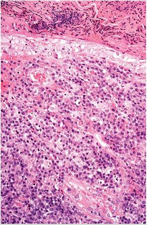
FIG. 6.53. Skin from the muzzle of a laboratory rabbit with rabbit syphilis. There is dense infiltration of the dermis with mixed leukocytes, degeneration of epidermis, and serous exudation on the surface. Source: A. Strom, University of California, Davis, CA.
Reproduced with permission from A. Strom.Diagnosis
Scrapings from lesions, with wet mount preparation and dark-field examination, is the recommended method for confirming the diagnosis. Demonstration of the spirochetes by silver staining of lesions in histology sections may be used (Fig. 6.54). Serology is a reliable diagnostic procedure. Tests available include the demonstration of plasma reagin antibody and the fluorescent treponemal antigen test. Differential diagnoses include moist dermatitis, Pasteurella infections of the external genitalia, and traumatic lesions.
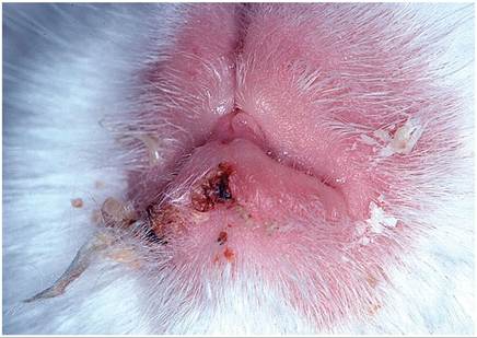
FIG. 6.52. Erosive chelitis in a laboratory rabbit infected with Treponema paraluis-cuniculi. The muzzle region is a frequent site for rabbit syphilis lesions.
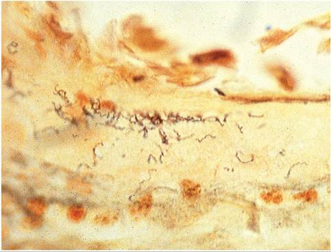
FIG. 6.54. Rabbit syphilis lesion, depicting numerous spirochetes within the surface exudate (Warthin-Starry stain).
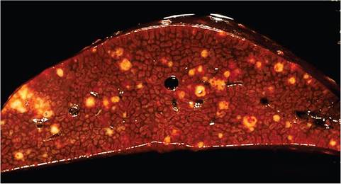
FIG. 6.55. Multifocal caseation necrosis in the liver of a rabbit naturally infected with Yersinia pseudotuberculosis. (Courtesy D. Imai.)
Yersinia pestis Infection: Plague
Wild rabbits are among many wild species that may become infected with Yersinia pestis, and transmission to humans from infected Sylvilagus spp. rabbits has been documented on multiple occasions. Infection of domestic rabbits is exceedingly rare. The bacterium causes septicemic disease in infected animals.
Yersinia pseudotuberculosis Infection: Pseudotuberculosis, Yersiniosis
Infection of domestic rabbits with Y. pseudotuberculosis is rare, but infection may be common in some populations of hares. It is among the most important lethal infections in wild hares in Europe. The bacterium is usually transmitted by the ingestion of food or water contaminated by birds and rodents. Lesions are characterized by focal granulomatous lesions in the intestine, with foci of caseation necrosis in the liver (Fig 6.55), spleen, lymph nodes, and reproductive tract.