INTRODUCTION
The Lentivirus genus was named to connote the slow and inexorable progression of the degenerative diseases its members cause in a number of mammalian species. The median time to progression to acquired immunodeficiency syndrome (AIDS) after primary HIV (human immunodeficiency virus)-1 infection in humans, for example, is approximately 10 years.
In other instances, however, a state of mutually benign accommodation seems to evolve, in which lentiviruses persist and replicate while causing no ill effects in host animals. The HIV-1 pandemic illustrates the pathogenicity that may ensue when a lentivirus has recently undergone cross-species transmission and remains unchecked by evolutionary coadaptation between parasite and host. Another biologically central issue, one that the prefix lenti belies, is the view we now have of HIV-1 replication and turnover in the body, where the process is anything but slow. Rather, a highly dynamic replication process is apparent, and the amount of HIV-1 circulating in plasma correlates very well with disease progression. This chapter will present an overview of these and other selected aspects of HIV-1 virology. The intent is to introduce the basic molecular biology of HIV-1 within a comparative lentiviral framework as a basis for considering the complex question that is the theme of the succeeding chapters in this volume: how do cells die from HIV-1 infection?Lentiviral biology and pathogenesis
Lentiviruses were the first retroviruses associated with disease and were among the first filterable disease agents identified.1 Three groups of lentiviruses infect primates, ungulates, and felines,
TABLE 1.1
Lentiviruses: Classification and Disease
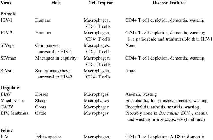
CD4+ T, CD8+ T, cat; asymptomatic in numerous other Felidae
and B cells
respectively (Table 1.1).
The term “slow virus” was first applied by Bjorn Sigurdsson in the 1950s during his studies of a maedi-visna virus (MVV) epidemic that illustrates well the potentially severe effects of introducing lentiviruses into naive populations or species.2 Importation of asymptomatic sheep from the European continent to Iceland in 1933 led to the subsequent death of more than 100,000 Icelandic sheep over several decades from MVV, which causes a pneumonic disease (maedi in Icelandic)3 and chronic encephalitis (visna).4,5 Both equine infectious anemia virus (EIAV), the disease agent described by Vallee and Carre in 1904,1 and MVV were studied as model slow viruses before primate lentiviruses were discovered.2-10 Feline immunodeficiency virus (FIV) and bovine lentiviruses (bovine immunodeficiency virus [BIV] and Jembrana disease virus) were isolated and characterized in the post-AIDS era.11-13Lentiviruses differ in a number of respects from other retroviruses. Among the most striking difference is their ability to replicate in terminally differentiated, nondividing cells.14,15 Recognition of this signature property inspired development of replication-defective vectors capable of permanent transgene integration in diverse nondividing cells of relevance to gene therapy.16-18 All lentiviruses infect nondividing tissue macrophages, which are the principal reservoirs in vivo for the ungulate lentiviruses. For example, the primary pathology caused by EIAV is a cyclical hemolytic anemia caused by antigen-antibody complexes that bind to the surfaces of erythrocytes; however, the primary EIAV producer cell is the macrophage.19,20 Lentiviral tropism is determined by receptor utilization. (See early events below.) FIV and the primate lentiviruses have evolved additional tropisms for lymphocytes. FIV has the broadest tropism, as it infects B cells and CD8+ T cells in addition to macrophages and CD4+ T cells.21 HIV-1 infects CD4+ T cells and macrophages and glial cells, although a variety of other cell types have been reported to be infected to a lesser extent in vivo.
More on the topic INTRODUCTION:
- Introduction
- Introduction
- Introduction
- Introduction
- Theory and Practice
- Hare C., Neo D. (eds.). Trade Finance: Technology, Innovation and Documentary Credit. Oxford University Press,2021. — 417 p., 2021
- AVIAN CHOLERA
- Barger A.M., MacNeill A.L. (Eds.). Small Animal Cytologic Diagnosis: Canine and Feline Disease. CRC Press,2024. — 536 p., 2024
- Harker C., Horschelmann K. (Eds.). Conflict, Violence and Peace. Springer,2017. — 456 p., 2017
- Abrams Peter A.. Competition Theory in Ecology. Oxford University Press,2022. — 336 p., 2022