INTRODUCTION
Depletion of peripheral CD4 T lymphocytes is a hallmark of HIV infection and is the result of many pathogenic mechanisms. The very high viral load observed during HIV primary infection leads to high levels of CD4 T cell activation and infection, which results in their elimination by apoptosis.
This cell loss must be compensated for by the production of naive T cells by the thymus. Unfortunately, thymic function is altered in the presence of HIV, and thymic output is reduced, which contributes to the reduction in peripheral CD4 lymphocyte pool. The apoptosis rate of thymocytes increases in HIV-infected patients, due to the pathogenic effect of HIV proteins, of HIV replication in thymocytes, and of perturbations in cytokine levels that result from the hyperactivation of the immune system. This review will focus on the alteration of T cell precursor survival in HIV-infected individuals as well as the molecular mechanisms involved in programmed cell death induced by HIV and that lead to the pathogenic effect of HIV on thymic function.the Apoptotic machinery in thymocytes
As in most mammalian cells, there are two main pathways that lead to programmed cell death (apoptosis) in thymocytes (Figure 20.1). The extrinsic pathway is triggered by the aggregation of death receptors (DRs), such as Fas/CD95, TNFR1/CD120a, or TNF-related apoptosis-inducing ligand (TRAIL) receptor/DR5, and leads to the activation of a caspase cascade that includes caspase- 8, caspase-3, -6, and -7.1 Apoptosis triggered by DRs is dependent on the function of Fas-associated protein with death domain (FADD), an adaptor protein required for caspase-8 recruitment to DR, and is inhibited by FLICE inhibitory protein (FLIP), a protein homologous to caspase-8 but lacking its proteolytic activity. The other apoptotic pathway is activated after DNA damage and triggers p53 activation, cytochrome c release from the mitochondria, formation of the apoptosome containing
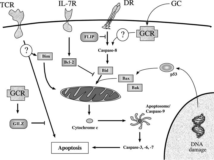
FIGURE 20.1 Molecular mechanisms involved in thymocyte cell death.
Thymocyte apoptosis can be triggered through the extrinsic pathway following death receptor (DR) engagement, the intrinsic pathway induced by DNA damage, the presence of glucocorticoids (GC), or T cell receptor (TCR) cross-linking by a high-affinity ligand (negative selection). GC can also antagonize TCR-induced apoptosis by increasing the expression of the GC-induced leucine zipper (GILZ) protein. Most of these apoptotic stimuli lead to cytochrome c release from the mitochondria to the cytosol and the activation of downstream caspases (caspase-3, -6, and -7). IL-7 also protects thymocytes from apoptosis by increasing levels of Bcl-2 expression.caspase-9, and subsequent activation of effector caspases (caspase-3, -6, and -7). This mechanism, called the intrinsic pathway, is inhibited by the antiapoptotic members of the Bcl-2 family, such as Bcl-2, Bcl-xL, or Bfl-1, whereas the proapoptotic members Bax, Bak, Bid, or Bad are required for cytochrome c release, subsequent caspase-9 activation, and cell death.2 Both pathways are functional in murine thymocytes that can be killed after anti-Fas stimulation3 or DNA damage4 in vitro. Caspase inhibitors block cell death induced through both pathways.
Glucocorticoids (GCs) also trigger massive cell death among double-positive (DP) thymocytes, through a caspase-dependent pathway.5,6 Bcl-2 and Bcl-xL overexpression protects DP thymocytes from GC-mediated killing.7,8 The GC receptor activates caspase-8 through a mechanism that is not well understood, which results in the release of cytochrome c from the mitochondria and caspase-9 activation (Figure 20.1).9 Paradoxically, GCs are also able to antagonize apoptosis after T cell receptor (TCR) cross-linking.10 The presence of GCs triggers the expression of GC-induced leucine zipper (GILZ),11 which inhibits TCR-mediated mitogen activated protein kinase (MAPK) activation12 and AP-1 and NF-κB activity.13,14 The role of GCs in thymic development is not completely understood, because positive and negative selection are unaffected in GC receptordeficient mice.15 Furthermore, although thymocytes can be killed in vitro by exposure to any of these stimuli, the signal that will determine whether a T cell progenitor undergoes apoptosis in the thymus is highly dependent on the recombination events that lead to the production of a functional TCR, as described below.