OUTSIDE INFLUENCES OR AN INSIDE JOB—THE EXTRINSIC AND INTRINSIC PATHWAYS
There are two general pathways of apoptosis (and a seemingly infinite number of variations). A simple distinction by intrinsic and extrinsic induction may be made to differentiate the two.
Both of these mechanisms use caspases to bring about the endpoints of death, but each relies on different initiators and engages the death pathway by different means.The extrinsic pathway is triggered by receptors at the cell surface.13,14 Receptor-mediated apoptosis is characterized by engagement of a death receptor from the tumor necrosis factor (TNF) family of transmembrane receptors, including Fas (CD95), TNF-R1, and DR4/5, by their corresponding ligands, Fas-ligand (CD95L; CD178), TNFα, and TRAIL, respectively. For a comprehensive review of the TNF and TNF-R family of proteins, see Locksley, Killeen, and Lenardo.13 Perhaps the simplest example is the pathway engaged by Fas by interaction with Fas-ligand. Receptor engagement results in recruitment of the adaptor protein FADD (Fas-associated death domain), which binds Fas by intermolecular interactions between death domains of Fas and FADD. Caspase-8 (or caspase-10) is then recruited to this complex to form what is called the DISC (deathinducing signaling complex). This pathway is exemplified with Fas in Figure 5.2. TNF-R1 was recently shown to activate caspase-8 by a two-step mechanism that includes recruitment of TRADD (TNF receptor-associated protein with DD) in a multimolecular complex leading to internalization of the DISC before recruitment of FADD and caspase-8 activation.15
Receptor-mediated apoptosis was implicated in the regulation of peripheral lymphocytes when it was discovered that the defect responsible for the lpr (lymphoproliferative) and gld (generalized
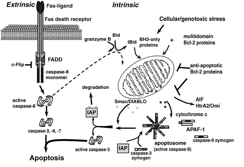
FIGURE 5.2 Overview of extrinsic and intrinsic apoptosis pathways.
The extrinsic is shown with Fas as a representative member of the TNF receptor family of death receptors. Fas-mediated signaling to death is perhaps the simplest of the known death receptor-mediated pathways.lymphoproliferative disorder) strains of mice were caused by mutations in Fas and Fas-ligand genes, respectively.14’16’17 Mutations in the corresponding human Fas and Fas-ligand counterparts have been implicated in autoimmune lymphoproliferative syndrome (ALPS) type Ia and Ib, respectively.
The intrinsic pathway is governed by the release of proapoptotic mitochondrial proteins including cytochrome c, Smac/DIABLO, EndoG, HtrA2∕Omi, and AIF.18 After release from the mitochondria, cytochrome c associates with the cytosolic adaptor protein, apoptotic protease activating factor (Apaf-1), along with dATP to recruit procaspase-9 into a macromolecular complex known as the apoptosome. Activation of the initiator caspase-9 then results in cleavage of caspase-3, but this is not yet sufficient to cause activation of the executioner. Smac/DIABLO, another of the mitochondrial proteins released along with cytochrome c, suppresses the inhibition of caspase-3 by XIAP (X-linked inhibitor of apoptosis protein [IAP]).19’20 De-repression of XIAP by Smac/DIABLO allows for release of active caspase-3 protease from the apoptosome into the cytosol, where it drives proteolytic degradation of the apoptotic cell.
In addition to cytochrome c, other mitochondrial proteins have been assigned apoptotic func- tions.18 EndoG is a mitochondrial endonuclease that, when released, has a demonstrated activity of chromosomal DNA digestion that does not require the involvement of caspases.21 The serine protease HtrA2/Omi was originally described as an IAP-antagonist with an effect similar to Smac/DIABLO in its ability to relieve IAP-induced repression of caspases. The IAP-inhibitory function of HtrA2/Omi is not dependent on its protease activity, which suggests an additional physiological role.
Recently, a role for HtrA2∕Omi as a sensor for misfolded mitochondrial proteins was proposed, providing another example of an apoptotic protein also having an important survival function.22 Similarly, AIF (apoptosis-inducing factor) is a resident flavoprotein in the mitochondrial intermembrane space that is capable of inducing chromatin condensation and degradation of chromosomal DNA into large fragments. Knockout of the AIF gene yielded a profound phenotype resulting in death during early cavitation of embryoid bodies, suggesting a key survival role very early in embryogenesis, perhaps throughout phylogeny.23The intrinsic pathway is regulated by members of the Bcl-2 protein family. Permeabilization of the mitochondrial membrane and release of apoptogenic proteins are inhibited by the pro-survival members of this family: Bcl-2, Bcl-xL, Mcl-1, A1, and Bcl-w.24 Bcl-2, the prototypic family member, was discovered in a clinical model of B cell lymphoma in which the Bcl-2 gene translocated downstream of the immunoglobulin heavy-chain promoter becomes grossly overexpressed.25 Dys- regulated overexpression of Bcl-2 in this scenario protects immature B cells from negative selection and results in subsequent emergence of follicular lymphomas.
The release of mitochondrial proteins is mediated by two classes of proapoptotic members of the Bcl-2 family, the BH3-only proteins in concert with the multidomain family members.26,27 The BH3-only proteins (including Bid, Bim, Bmf, Bik, BNip3, Puma, and Noxa) are viewed as cellular sensors that transmit a distress signal to effect the death program. Mitochondria form a central transmission point of the death signal, where BH3-only proteins interact with a corresponding member of the multidomain group (Bax, Bak, Bad). The biochemical mechanisms by which the proapoptotic members interact are reviewed in Scorrano and Korsmeyer.27