Complications of the puerperium
Infection
The risk of infection is ever present for women after childbirth and some infections have the potential for severe disease and even death. Events during labour and delivery, such as vaginal examinations, prolonged rupture of the membranes, perineal injuries, instrumental delivery, and manual removal of the placenta, all have the potential for associated infectious complications.
The presence of devitalized tissues, blood clot, or haematoma anywhere in the genital tract can foster development of infection. For these reasons a high level of suspicion and low threshold for treatment are important in the safe management of puerperal infection.Vaginal and perineal injuries
Perineal injury, especially if there has been some delay in repair, may become infected. This is particularly so where there has been a third- or fourth-degree injury or where an associated haematoma is present or has been evacuated (Figure 35.4). The perineum can be heavily soiled after birth so attention to cleansing and aseptic technique during suturing are important preventive measures. Where anal sphincter or rectal mucosal injuries have been treated, a case can be made for use of antibiotics at the time of repair and in the immediate postpartum period although evidence to guide practice can be difficult to obtain (22). Advice regarding perineal care should be provided, for example, twice-daily showers and avoidance of traumatic drying of the skin. Although perineal infection is relatively uncommon, such infections are important as they are commonly associated with breakdown of the suture line, a particularly unpleasant complication for women (23). Careful surveillance for healing, and early treatment of suspected infection are important principles of care. Perineal infections are usually polymicrobial with facultative and anaerobic species so broad-spectrum antibiotics including metronidazole will usually be required.
Caesarean wound infection and necrotizing fasciitis
As caesarean section rates increase, infection of abdominal incisions has become a more common complication. Some degree of infection of the caesarean section incision occurs in about 10% of cases, and is more common after emergency caesarean section (24). The majority of wound infections are diagnosed after discharge from hospital and are, fortunately, relatively mild. Obesity is a major risk factor for caesarean wound infection and is becoming more common in the obstetric population (25). The use of perioperative antibiotics at the time of caesarean section, and swabbing the vagina with antiseptic solution prior to delivery have been shown to reduce the risk of infection in the incision (26). Despite the best technique and management, caesarean wound infection still occurs and management must
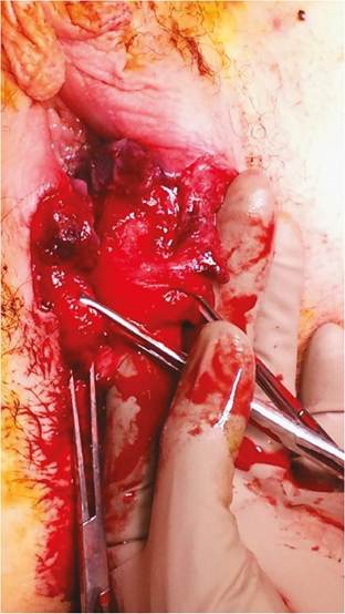
Figure 35.4 Third-degree perineal tear, with complete division of the internal and external anal sphincters. Careful, precise repair of such injuries and close postpartum surveillance and management are the keys to a good long-term outcome from such injuries.
be prompt and careful. Where cellulitis is the primary manifestation, use of intravenous antibiotics is appropriate. However, if there is evidence of purulent discharge or a deeper collection then drainage and debridement, if necessary, should be undertaken without delay (27).
Necrotizing fasciitis following caesarean section is a rare but potentially devastating complication. It occurs when there is a rapidly spreading gangrenous infection of the deep tissues of the abdominal wall, typically polymicrobial in character with Clostridium and group A Streptococcus (GAS). The presentation may be delayed up to a week or more following birth, with patients reporting increasingly severe pain and debility. Oedema and discolouration of the skin may be seen, and imaging may show gas collections in the abdominal wall.
A diagnosis of necrotizing fasciitis following caesarean section is an emergency situation, as the condition is life-threatening and demands urgent action. A combination of high-dose antibiotic therapy and aggressive surgical debridement is necessary, undertaken with a multidisciplinary approach (27).Mastitis and breast abscess
Mastitis is a relatively common complication of birth, affecting up to one-third of all woman at some point during lactation with the majority of cases occurring within the puerperium (28). Fortunately, the severe complication of mastitis—breast abscess—is rare and affects less than 0.5% of breastfeeding women (29). Some women are prone to mastitis and have repeated episodes, although for most women mastitis is a single event during lactation.
Mastitis typically occurs in women with predisposing factors such as nipple trauma and difficulties in feeding, and possibly reduced frequency of feeds. Maternal fatigue and primiparity may increase the risk, as does a history of past episodes of mastitis. The commonest pathogen isolated is Staphylococcus aureus, followed by various species of streptococci (28).
The pathophysiological mechanism underlying acute mastitis is milk stasis. Milk stasis appears to have an inflammatory effect on the breast tissues, and is associated with maternal fever and breast engorgement. This may be due to the presence of inflammatory mediators in human milk, and a range of proteins not found elsewhere in the body (28). The risk of milk stasis can be reduced by early exclusive breastfeeding, not restricting access to the breast for babies, and support and encouragement of comfortable and functional motherbaby positioning for feeds and correct attachment to the nipple to minimize traumatic injury.
Mastitis can occur very suddenly following a brief prodrome of fever, rigors, and feelings of an influenza-like illness with generalized pain and malaise. Because the infection spreads from the ductal system into the connective tissue, the affected breast becomes painful and may develop an angry erythema that spreads out from the affected area.
There is seldom time for breast milk culture and microscopy, and broad-spectrum antibiotics and adequate analgesia should be instituted rapidly. Broad-spectrum antibiotics with anti- staphylococcal sensitivity such as oral cephalosporins are usually indicated. Women will need analgesics with antipyretic properties, such as paracetamol, and possibly non-steroidal anti-inflammatory medication.It is important that milk stasis is minimized in the affected breast so the baby should continue to feed from the affected side, ideally as the first side to promote effective suckling. Alternatively, a breast pump may be used to empty the breast and this can also be useful after a feed to ensure that the affected breast has been completely emptied. Time spent with the mother to assess and, if necessary, adjust feeding technique or timing is very useful and may help reduce the risk of further episodes. Once antibiotic therapy has been instituted, it should be continued for at least 10 days to reduce the risk of relapse. The support of maternity staff and the woman's family are very important during treatment of mastitis as it is very unpleasant for the women and can be extremely debilitating and stressful.
Incomplete treatment of mastitis may lead to development of a breast abscess. In this case, the woman fails to improve with standard management and the breast may become fluctuant with the woman experiencing spiking fevers. Imaging may be required to demonstrate the presence of an abscess, and where therapy with high-dose intravenous antibiotics does not lead to an improvement in the clinical picture, rarely incision and drainage will be necessary.
Urinary tract infection
Urinary infection is a common cause of fever in the early postpartum period and although women may report urinary frequency and dysuria these symptoms are not invariable and symptoms may be subtle. A high index of suspicion should be maintained where there is a history of catheterization during labour or afterwards, and where there has been perineal or vaginal trauma and swelling.
Accurate diagnosis can be difficult due to contamination with lochia, and the presence of white cells in the urine commonly results from bladder trauma during birth.The commonest organism is Escherichia coli, but other pathogens include Gram-negative species such as Klebsiella and Enterobacter, as well as streptococci (30). Where the woman is febrile and has flank pain, treatment with intravenous antibiotics should be commenced promptly. Due to the high prevalence of resistant bacteria, a broad-spectrum cephalosporin would usually be the treatment of choice. In a clinical setting suspicious for more severe infection, an aminoglycoside should be added to the regimen. Failure to respond to treatment within 2 or 3 days should prompt investigation for underlying complicating factors such as voiding dysfunction, obstruction, or other pathologies such as a renal abnormality or calculus.
Endometritis and retained products of conception
Postpartum endometritis complicates as many as 5% of vaginal births, and a higher proportion of caesarean deliveries (30). The typical presentation is with lower abdominal pain accompanied by malodourous vaginal lochia. Predisposing factors include a history of prior bacterial vaginosis, a prolonged labour, intrapartum fever, and conditions such as diabetes and anaemia. Examination will typically reveal fever and tachycardia with a tender uterus, but absence of fever does not exclude the presence of endometritis. The presence of an elevated white cell count and raised inflammatory markers, such as C-reactive protein, are non-specific but lend support to the clinical diagnosis. Similarly, the results of microscopy and culture of vaginal swabs can be helpful but do not necessarily reflect conditions within the endometrial cavity. Imaging is useful to exclude retained products of conception or pelvic abscess.
Infection is typically polymicrobial with anaerobes and genital mycoplasmas, and occasionally Chlamydia species when the onset of clinical infection is delayed (30).
Obtaining accurate cultures from the endometrium can be difficult, and is often delayed by several days—pathogens are uncommonly identified in blood cultures or urine specimens. Where the clinical presentation suggests moderate severity with a picture of acute sepsis, purulent discharge, and pain, treatment should begin promptly and consist of fluid resuscitation and pain relief, as well as broad-spectrum antibiotics. Most women will improve quickly, and where there is no improvement over 2-3 days, investigations should aim to exclude pelvic thrombophlebitis or abscess, unsuspected infection elsewhere, or resistant strains (30).Group A Streptococcus
Systemic infection with GAS—puerperal sepsis—is the feared infection for the postnatal women, and there is some evidence that the incidence of GAS is increasing. Although infection can occur ante- natally, it is more common and potentially devastating after birth. GAS colonization is common, with as many as one-quarter of the population identified as asymptomatic carriers. The typical presentation is with high fever, rigors, and rapidly progressing malaise. In some cases, women will report a prodromal period with a sore throat or non- specific influenza-like illness. There may be symptoms or signs of endometritis, with vaginal discharge or bleeding and a tender lower abdomen and uterus.
Although many cases have no identifiable risk factors, retained products of conception, prolonged labour, and instrumental delivery may be present. Woman at social disadvantage and those with known immunosuppression or chronic disease may be at higher risk, but overtly healthy women can be affected. A high index of suspicion must be maintained and women who present with high fever and malaise in the postnatal period should have blood cultures, a urine specimen, and swabs taken with a specific request for M-typing of any GAS isolated from the specimens. Treatment should be aggressive and multidisciplinary with involvement of clinical microbiologists and intensive care teams. High-dose broad-spectrum antibiotics including penicillin and fluid resuscitation with full supportive care are commonly required.
Secondary postpartum haemorrhage
Bloody-stained loss is common in the postnatal period, but secondary haemorrhage is said to occur where there is increasingly heavy frank blood loss beginning more than 24 hours after birth. Unlike primary postpartum haemorrhage (PPH), which is often defined in volume terms (such as 500 mL or more, or 1 L in other definitions), there is no agreed volume in definitions of secondary haemorrhage. Because of the lack of standardization it is difficult to provide a precise incidence rate for secondary PPH with studies estimating that between 0.5% and 1.5% of women will be affected (31). Although much less common than primary PPH, secondary PPH is still a source of morbidity and even maternal death.
The factors predisposing to secondary PPH are those that interfere with the normal involution of the uterus, such as retained placental tissue and membranes, secondary infection, and the presence of leiomyomata (fibroids). Secondary infection may be associated with prolonged prelabour rupture of the membranes, prolonged labour, emergency caesarean section, and manual removal of the placenta (31). Less commonly, secondary PPH occurs with undiagnosed placenta accreta, lower genital tract injuries (especially if there has been haematoma formation), complications of a caesarean section incision such as dehiscence, unrecognized vascular abnormalities such as arteriovenous malformations, and where a coagulopathy develops subsequently to the birth.
The management of secondary PPH should follow a standard approach, with early recognition and action the cornerstone of a good outcome. Although such bleeding is usually light, rarely it can be heavy and potentially catastrophic (32). Resuscitation should be prompt and aggressive as required, with a low threshold to use blood or blood products if early therapy with plasma expanders is unsuccessful. Fortunately, most women will be haemodynamically stable and time can be taken to establish a diagnosis and to tailor treatment to the clinical situation. Examination may reveal potentially helpful signs such as fever, abdominal tenderness, larger-than- expected uterine size, offensive discharge, or local perineal bleeding or haematoma.
Investigations should include a check of haemoglobin concentration and platelet count, with coagulation studies if there is watery loss and absent clotting, as well as grouping of blood in case transfusion is required. Vaginal swabs should be taken to establish whether infection is present, and to guide antibiotic therapy if required. Ultrasound examination is the cornerstone for diagnosis of retained tissues, although in the very early postpartum period interpretation of the findings require experience and judgement (31). Use of colour Doppler, if available, may help distinguish between retained placental tissue (which may retain a blood supply) and necrotic clot or decidua (33). While false-positive results from early postpartum ultrasound scanning may lead to unnecessary surgical intervention, imaging revealing an empty uterus can spare a woman anaesthesia and exploration of the uterus where it is not required.
The therapy for secondary PPH will depend upon the suspected cause. Uterotonic agents such as misoprostol (which can be administered orally or rectally) or prostaglandins (given intramuscularly) may be used to assist with uterine contraction. Although oxytocin is usually readily available, its effectiveness decreases the greater the time since birth. Since infection and resulting endometritis are likely to contribute in many episodes of secondary PPH, antibiotic treatment is commonly instituted (34). Typical bacteria isolated in the setting of secondary PPH include E. coli, Clostridium perfringens, Bacteroides, and streptococci (31). When there is either a clinical suspicion or direct ultrasound evidence for retained tissue or clot, and surgical evacuation is planned, antibiotic treatment may be commenced prior to the procedure. Where there is some clinical urgency, use of antibiotics at the time—usually with induction of anaesthesia—should be considered. Although protocols for antibiotic use will vary between institutions, the use of broad-spectrum antibiotics likely to cover the typical range of infective organisms may include penicillins, metronidazole, and gentamicin (31).
There is no single recommended method of uterine evacuation and manual exploration and evacuation, use of a wide-bore suction catheter, and sharp curettage have all been described (35). There is a greater chance of uterine perforation and injury when curettage is performed in the early postnatal period so care and precision are important, particularly in a setting of caesarean section. Fortunately, retained placental tissue is less likely following caesarean section. When infected retained tissues are evacuated surgically, there is an increased risk of uterine synechiae and Asherman syndrome although these are difficult to avoid and the risks must be balanced against continued bleeding and possible escalation of infection (36).
Voiding dysfunction
Voiding difficulty and associated urinary retention is a relatively common occurrence in the puerperium. Although definitions vary, estimates of the incidence of overt voiding dysfunction suggest about 5% of women are affected with a further 10% affected by less obvious dysfunction (37). The pathogenesis of voiding dysfunction is usually multifactorial, resulting from a combination of mechanical, neurological, and physiological processes. Progesterone has an inhibitory effect on smooth muscle tone and this may contribute to reduced detrusor muscle tone.
Voiding dysfunction has a range of clinical presentations, with a complete inability to pass urine in the most severe group. However, there is a spectrum of effect with some women remaining asymptomatic yet having a large postvoid residual bladder volume (38). A high index of clinical suspicion should be maintained in women with recognized risk factors: epidural and regional anaesthesia, primiparity, instrumental delivery, a large baby, and a long duration of labour (38). Women with sequelae of a traumatic vaginal birth, such as prominent vulval oedema, bruising, and tears in the anterior vulva and vagina near the urethra, require special care. Typical presentations include a frequent need for urination with small volumes passed, hesitancy or a need to strain with voiding, a slow or intermittent urinary stream, or urinary urgency. Some women with voiding dysfunction will report a lack of sensation to void.
Proactive surveillance and management are important to avoid short- and longer- term voiding problems. Vulval oedema and trauma should be managed with application of cold packs and selective use of anti-inflammatory medication, and effective, regular analgesia to reduce hesitancy due to pain. Women should be encouraged to ambulate to the toilet and be afforded privacy. Any woman who has not voided within 6 hours of birth should have her residual volumes measured, ideally by catheterization since the results of ultrasound bladder scanners may be affected by the postpartum uterus. Where the woman remains unable to void and has residual bladder volumes of more than 150 mL, an indwelling catheter should be left in place for 24-48 hours then another trial of void undertaken (38). If the voiding dysfunction persists, a catheter should be left in place for 1 week with antibiotic prophylaxis to reduce the risk of cystitis.
With prompt diagnosis and systematic management, most women with voiding dysfunction in the early postnatal period will recover completely and have no enduring difficulties. Although the long-term prognosis is excellent in virtually all cases, persistent voiding dysfunction can necessitate intermittent self- catheterization for weeks which is highly disruptive and predisposes to repeated urinary infections.
Constipation and haemorrhoids
Constipation is a commonly reported problem in the puerperium, affecting up to 20% of women. Although there is wide variation in the normal patterns of bowel function, bowel movements that occur less than three times a week with hard stool that is difficult to pass would generally be diagnosed as constipation. When constipation is diagnosed, careful assessment and management are required. Factors that can exacerbate constipation include opioid pain relief and iron supplements. Physical inactivity and depression are associated with constipation and conditions that make it painful to defecate, such as haemorrhoids and anal fissures, can lead to faecal impaction and sometimes overflow incontinence.
Management should be based on ensuring the woman has an adequate intake of fibre and fluid, and that defecation should not be delayed if there is an urge. Where these measures do not provide adequate relief, stepwise use of bulk-forming laxatives is usually the next step. Agents such as psyllium, ispaghula, and wheat dextrin are safe and effective, but may take a couple of days to work and need to be introduced gradually. Women should be warned about symptoms such as bloating and abdominal discomfort, and need to have a good fluid intake at the same time. If these measures are not successful then osmotic laxatives should be added in addition, either in the form of oral solutions or sometimes by the rectal route. It is uncommon for women still to have problems following these measures, but where success has still not been attained, further and more specialized assistance and advice may be required.
Haemorrhoids are common in pregnancy, and often undergo an acute exacerbation following birth. They result when the supporting tissues of the anal cushions fail, allowing prolapse of the cushions and venous engorgement. There is commonly associated venous thrombosis and a marked inflammatory response, making them very painful to endure (39). Haemorrhoids present as an uncomfortable lump or lumps at the anus, and can cause bleeding during or after defecation. There may be irritation or pruritus associated with faecal soiling, but acute pain is usually associated with either a large acute thrombosis, or with an associated anal fissure.
In general, the management of haemorrhoids in the postnatal period is conservative with the use of Sitz baths up to three times a day, and a pillow or ring cushion to sit on at other times. Women may need to use analgesic and anti-inflammatory creams or ointments before defecation. An important aspect of managing haemorrhoids is to attend to constipation. Women should be reassured that haemorrhoids will settle within a week or two of birth. Where there are continuing problems, the opinion of a general or colorectal surgeon should be sought.
Urinary incontinence
The prevalence of urinary incontinence for women in the postnatal period is surprisingly high, with pooled data from population studies suggesting that one woman in three will report episodes of involuntary urine loss (40). Of note, incontinence is twice as common in women who have had vaginal deliveries when compared to those whose birth was by caesarean section. Reporting urinary incontinence can be embarrassing for many women, so direct questioning may be necessary. In all cases urinary infection should be excluded and other pathology, such as pudendal nerve injury, should be sought. Women who describe incontinence should be managed sensitively in conjunction with an experienced physiotherapist. Therapeutic strategies include coaching in pelvic floor exercises, attention to general fitness, and optimization of weight, and in some cases bladder retraining.
Faecal incontinence
Faecal incontinence following birth is less common than urinary incontinence, but still affects up to 5% of women in the postnatal period, and this figure might be low due to under-reporting (41). The commonest cause is injury to the anal sphincter which complicates up to 3% of all vaginal births. Accurate diagnosis of anal sphincter injuries is important as it allows correct surgical repair and appropriate postnatal management. However, it is likely that many injuries are not recognized at the time of birth and even women who have undergone satisfactory repair are likely to report impairment of bowel control in the postnatal period (42). When there is a sensation of urgency and associated faecal incontinence it is likely that damage to the external anal sphincter has occurred. Injury to the internal sphincter typically presents as passive leakage of flatus or faeces.
There are other causes of these symptoms, including faecal loading of the bowel with overflow. This can be associated with use of narcotic pain relief medications, or where there is significant pain that inhibits normal defecation. When a sphincter injury has occurred, it is important to have frequent contact with the women in the postnatal period, and to provide advice and assessment and ongoing management. Assessment should include careful regular examination of the perineum, anus, and vagina to check that healing is progressing satisfactorily and to assess for asymmetry with voluntary contraction, to assess for reflex contraction with coughing, and to assess sphincter tone. At that same time, haemorrhoids or anal fissure can be excluded, or diagnosed if present and treated appropriately.
Women should be offered advice on diet and the importance of adequate fibre and fluid intake, and also coaching in pelvic floor exercises. One of the most important aspects of care is to offer emotional support, as caring for the newborn is stressful in itself without the added burden of uncertainly and lack of confidence that comes with faecal incontinence. Where women do not respond to conservative measures, referral for further assessment and possibly secondary repair should not be delayed.
Common breastfeeding difficulties
Breastfeeding is an important measure in promoting the health and well-being of children and for this reason should be supported and encouraged. However, almost one-third of women will report difficulties in breastfeeding and these usually fall into two broad categories—pain and a perception of low milk supply (43). Breast and nipple pain are an important symptom and the first condition to exclude is mastitis (discussed previously). Where mastitis is not present, poor attachment of the baby to the nipple is the commonest cause of pain. This can occur when the nipple is inverted and difficult for the baby to grasp, or where there is restricted jaw or tongue movement for the baby. In some cases the frenulum under the baby's tongue may be snipped with sterile scissors and this does not seem to cause undue distress for the baby and may improve attachment.
Where there is trauma to the nipple it is common for the area to be colonized with staphylococci, so use of a topical antibiotic may be of value. Use of purified lanolin has also been shown to be effective in assisting the nipples to heal (43). Where these measures do not provide relief, there has been a presumption that Candida infection of the nipples may be a factor and there is certainly some evidence that topical antifungals to both the nipples and the baby's mouth may help (44). Another possible cause is vasospasm, related to Raynaud's phenomenon. This presents with blanching or other colour changes to the nipple, and acute onset of pain radiating into the breast. It commonly occurs in women with a prior history of Raynaud's. The main precipitant is cold so avoidance of cold by covering the nipple immediately after feeds is important. In refractory cases use of the peripheral vasodilator nifedipine has been recommended, although at present there are few trials to confirm this (43).
Low milk supply is commonly reported but is a very difficult problem to define in practical terms. Continuing adequate supply depends upon regular removal of milk from the breasts, a normal hormonal milieu, and adequate breast tissue. Breast hypoplasia is relatively uncommon, and some women will have had breast reduction surgery, but the majority of women will have adequate breast tissue. In many cases the perception of low milk supply results from misinterpretation of feeding patterns or a concern that the breasts are ‘soft:'. If the baby is gaining weight satisfactorily, the milk supply is likely to be normal and sometimes reassurance is all that is required. A factor that has been demonstrated to contribute to continued successful breastfeeding is skin-to-skin contact after birth, and this should be encouraged (45). Where reduced milk supply is suspected, the use of manual compression of the breast while expressing milk may increase production, along with attention to effective attachment, increasing frequency of feeds, expressing after feeds, and ensuring that the baby is offered both sides (43). Although ‘galactogogues' such as domperidone or metoclopramide are commonly prescribed, there is little evidence that they are effective at this time.
Fistula
Obstetric fistula is a major public health problem in developing countries with an incidence rate of up to 1 in 500 births, of which 80% result from obstructed labour (46). A combination of sociocultural problems promote the occurrence of fistula, including mal- nourishment leading to skeletal maturation, and marriage at an early age with childbearing before growth of the pelvic bones is complete. In regions where these risk factors are common, there is often a lack of access to obstetric care and facilities for emergency birth by caesarean section (47). In the majority of cases the baby dies, doubling the tragedy for the woman.
Almost half of all women who develop obstetric fistulae are primigravid, and there is often a delay of years between the labour and the seeking of medical attention (48). Findings can be complex and include vesico-, urethro-, and Ureterovaginal fistula often with loss of bladder tissue due to pressure necrosis. Rectovaginal fistula formation is also common and is associated with pelvic inflammatory disease and secondary infertility. With these terrible injuries comes chronic skin excoriation due to maceration by urine or faeces. Unsurprisingly, women with fistulae commonly find themselves cast out and abandoned by their families, driving them into social isolation, divorce, poverty, malnutrition, and severe mental health problems sometimes culminating in suicide (47).
More on the topic Complications of the puerperium:
- Complications of the puerperium
- Arulkumaran S., Ledger W., Denny L., Doumouchtsis S. (eds.). Oxford Textbook of Obstetrics and Gynaecology. Oxford University Press,2020. — 928 p., 2020
- Detailed contents
- Treatment postpartum-immediate and long term
- REFERENCES
- Cervical intraepithelial neoplasia
- 5 Normal Labor and Delivery, Operative Delivery, and Malpresentations
- Anticoagulants in pregnancy
- Chapter 11 Postpartum Care
- REFERENCES