Fertilization and implantation
Gametogenesis
The full maturation of spermatozoa takes approximately 64-70 days. FSH causes stimulation of spermatogenesis and LH is responsible for stimulation of Leydig cells and testosterone production.
A large number of spermatogonia are produced by mitosis after puberty and are converted to spermatocytes in the testis. Following the first meiotic division, spermatozoa are released into the seminiferous tubules and then into the vas deferens. The second meiotic division is then completed (Figure 1.6).
Follicular development is characterized by enlargement of the ovum with aggregation of stromal cells to form the thecal cells. When a dominant follicle is selected, the innermost layers of granulosa cells become adherent to the ovum and form the corona radiata. A layer of gelatinous material around the ovum forms the zona pellucida. The follicle enlarges and bulges through the surface of the ovary and is released at the time of ovulation. The granulosa and the theca internal cells undergo luteinization. Formation of the corpus luteum occurs approximately 7 days after ovulation. Unless implantation occurs, it subsequently regresses.
Sperm transport
Once sperms arrive near the cervical os, sperm migration into the cervical mucous occurs with a rate normally of 6 mm/min. Motile
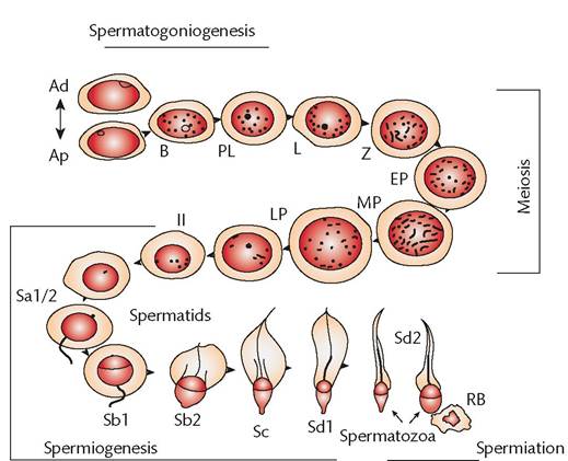
Figure 1.6 Schematic representation of the germ cell types and their development path during human spermatogenic process. Ad, A darkspermatogonium; Ap, A pale-spermatogonium; B, B-spermatogonium; EP (early), MP (mid), and LP (late), pachytene spermatocyte; II, secondary spermatocyte; L, leptotene spermatocytes; M, mitochondria; PL, preleptotene spermatocytes; RB, residual body; Sa-Sd2, steps of spermatid differentiation (Sd2 spermatids are the mature testicular sperm).
The developmental process from spermatogonium to formation of testicular sperm is considered to require at least 64 days (33-35).Reproduced from C. Marc Luetjens and Gerhard F. Weinbauer, The male gamete: spermatogenesis, maturation, function, in: Oxford Textbook of Endocrinology and Diabetes 2e (eds: John Wass et al.), Oxford University Press, 2011, with permission from Oxford University Press.
spermatozoa reach the uterine cavity and subsequently the fimbrial end of the fallopian tube.
Capacitation and fertilization
Capacitation is the functional maturation of the spermatozoon. It takes place once sperm passes through the epididymis and seminal vesicles. This process continues in the uterus or fallopian tube. Capacitation allows penetration of the zona pellucida by the sperm. Enzymes such as beta-amylase or beta-glucuronidase may act on the membranes of spermatozoa and facilitate sperm penetration. The capacitation process also involves modifications of membrane lipids, loss of cholesterol from plasma membrane, activation of the cyclic adenosine monophosphate/protein kinase A (cAMP/ PKA) pathway, increases in calcium (Ca2+) uptake and pH, hyperpolarization of membrane potential, and tyrosine phosphorylation (10).
The process of fertilization involves the union of the ovum and spermatozoon. When a spermatozoon reaches the cumulus around the ovum, the acrosome reaction is initiated. The outer acrosomal membrane fuses with the plasma membrane surrounding the spermatozoon and lytic enzymes are released. This facilitates the penetration of the oocyte membrane. The sperm head fuses with the oocyte plasma membrane and by phagocytosis the sperm head and mid piece are engulfed into the oocyte. The tail piece is left outside the cell membrane of the oocyte.
The sperm head forms the male pronucleus and with the female pronucleus they form the zygote. After disintegration of the membranes of the pronuclei, fusion of the male and female chromosomes occurs.
This is called syngamy and is followed by the first cleavage division. After a series of divisions and at the 16-cell stage, a solid ball of cells called blastomeres forms within the zona pellucida. This is known as a morula. A fluid-filled cavity develops within the morula to form the blastocyst.Implantation
Thirty-six hours after fertilization, the conceptus is transported through the fallopian tube and reaches the uterine cavity approximately 4 days later. The secretory endometrium is receptive to implantation. The second mitotic division of the oocyte is completed after fertilization and is followed by extrusion of the second polar body.
Six days after ovulation, the embryo becomes attached to the mid portion of the uterine cavity. By the seventh day, the blastocyst lies deep in the endometrium.
Physiology of coitus
The sexual response cycle described by Masters and Johnson has four phases in males and females. These phases are excitation, plateau, orgasmic, and resolution (Figure 1.7) (11, 12).
In the male, the excitation phase is associated with increased blood flow to the genitals and compression of venous channels of the penis resulting in erection. In the plateau phase, the penis remains in erection and the testes increase in size. Secretion of clear fluid may appear at the urethral meatus. In the orgasmic phase, reflex contractions of the bulbospongiosus (formerly bulbocavernosus) and ischiocavernosus muscles are followed by ejaculation of semen in spurts. During the resolution phase,
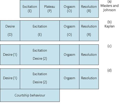
Figure 1.7 The development of the human sexual response model from (a) the original excitation, plateau, orgasmic, and resolution model of Masters and Johnson (11) through (b) the desire, excitation, orgasmic, and resolution model of Kaplan (12) to (c) the proposed modification with desire phase 1 (before initiation of the excitation phase and desire phase 2 during excitation phase) and finally (d) with added courtship behaviour.
Reproduced from Roy J. Levin, Normal sexual function, in: New Oxford Textbook of Psychiatry (eds. Michael Gelder, Nancy Andreasen, Juan Lopez-Ibor, and John Geddes), Oxford University Press 2012, with permission of Oxford University Press.
In the female, the excitation phase involves erection of the clitoris and swelling of the labia minora and vagina, vaginal lubrication, and nipple erection. The orgasmic phase is associated with narrowing of the vaginal introitus and contractions of the pelvic floor muscles. The plateau phase may be sustained in females and result in multiple orgasms. Following orgasm, congestion of the pelvic organs resolves rapidly.