Embryology
Early embryo development
Cells differentiate into an outer layer, the trophoblast, and an inner cell mass, which will give rise to the embryo proper, the amnion, yolk sac, and allantois.
Only two layers of cells intervene between the amniotic sac and yolk sac (Figure 1.8).The layers of cells adjacent to the amniotic sac form the embryonic ectoderm. Ectodermal tissues of the fetus develop from these cells including skin, its appendages, neural tube, and its derivatives (brain, spinal cord, autonomic ganglia, and adrenal medulla). Cells adjacent to the yolk sac form the embryonic endoderm. Endodermal tissues include lining of the gut, epithelial cells of thyroid, parathyroid, trachea, lungs, liver, and pancreas. Between ectoderm and endoderm, a third layer of cells develops mainly from ectodermal proliferation. This middle layer forms the mesoderm. Mesodermal tissues are bones, muscles, cartilage, and subcutaneous tissues of the skin.
Organogenesis
Ectoderm, mesoderm, and endoderm initially take the form of a circular sandwich. Disproportionate growth of ectoderm results in elongation of the embryonic plate into an oval form. Each end of this plate curves, forming the head and tail folds. The amniotic sac enlarges and completely surrounds the developing embryo and the yolk sac. On the dorsal aspect of the ectoderm, a groove appears from the middle of the head to the tail and changes into the neural tube from which the nervous system develops.
Mesoderm starts to grow laterally and gives rise to the paraxial mesoderm, the intermediate cell mass, and the lateral plate mesoderm (Figure 1.9).
Endoderm grows first laterally and then ventrally to form the gut.
The lateral plate of the mesoderm divides into somatopleure, which remains adjacent to ectoderm, and the splanchnopleure, which grows around the developing gut. The space between the somatopleure and splanchnopleure form the coelomic cavity.
This later becomes the pleural and peritoneal cavities.The paraxial mesoderm develops into vertebrae, dura matter, and muscles of the body wall. The intermediate cell mass grows ventrally into the coelomic cavity and forms the urogenital system. See Figure 1.10.
Development of the genital organs
Genital and urinary systems arise from the intermediate mesoderm. The pronephros appears first and quickly disintegrates. At the caudal end of pronephros, the mesonephric duct (Wolffian duct) develops
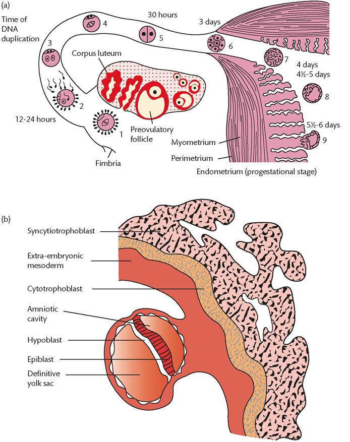
Figure 1.8 (a) Events of the first 6 days of development of a human embryo. 1: oocyte immediately after ovulation; 2: fertilization 12-24 h later results in the zygote; 3: zygote contains male and female pronuclei; 4: first mitotic division; 5: two-cell stage; 6: 3-day morula made up of up to 16 blastomeres; 7: morula stage (16-32 blastomeres) reaches the uterine lining; 8: early blastocyst; 9: implantation occurs at around day 6. (b) The site of implantation at the end of the second week.
Reproduced from Robert Wilkins, Simon Cross, Ian Megson, and David Meredith, Reproduction and development, Oxford Handbook of Medical Sciences, 2011, with permission from Oxford University Press.
and passes down the body to reach the cloaca. The mesonephros develops as a bulge in the dorsal wall of the coelom in the thoracic and lumbar regions. Two important structures appear on the coelomic surface of the mesonephros: (a) the genital ridge from which the gonad will form and (b) the paramesonephric (Mullerian) duct. The paramesonephric duct appears as a groove on the lateral aspect of the coelom and then becomes a tube. See Figures 1.11-1.14.
Placental development
Following implantation, the trophoblast completely surrounds the embryo in the form of proliferating cytotrophoblast and a syncytial layer. During the first trimester, a subset of trophoblast cells, the extravillous trophoblast (EVT) cells, become invasive and grow through the outer syncytium into the decidua where they invade maternal spiral arterioles.
The trophoblast shell begins to break open, allowing maternal blood to enter the primitive intervillous space where it is utilized by the developing villous placenta and fetus. See Figure 1.15.Development of membranes and amniotic fluid
The embryonic disc lies between the amniotic cavity and the primary yolk sac. The amniotic cavity develops between the embryonic ectoderm and cytotrophoblast. By the 12th postovulatory day, the base of this cavity is formed by embryonic ectoderm and the walls and roof are formed by the cytotrophoblast. Amniotic fluid is initially formed from the primitive cells around the amniotic vesicle.
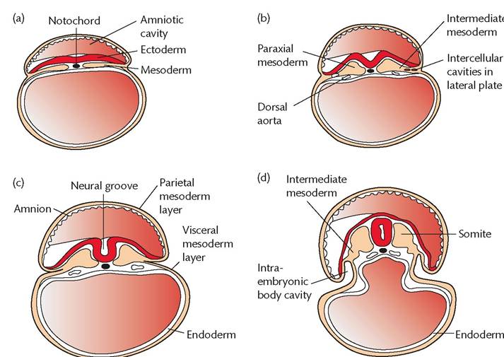
Figure 1.9 Transverse sections showing development of the mesodermal germ layer at days 17 (a), 19 (b), 20 (c), and 21 (d).The thin mesodermal sheet gives rise to paraxial mesoderm (future somites), intermediate mesoderm (future excretory units), and lateral plate, which is split into parietal and visceral mesoderm layers lining the intraembryonic cavity.
Reproduced from Robert Wilkins, Simon Cross, Ian Megson, and David Meredith, Reproduction and development, Oxford Handbook of Medical Sciences, 2011, with permission from Oxford University Press.
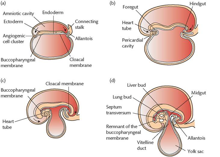
Figure 1.10 Sagittal midline sections of embryos at various stages of development demonstrating cephalocaudal folding and its effect on position of the endoderm-lined cavity. Presomite embryo (a), seven-somite embryo (b), 14-somite embryo (c), and 1-month embryo (d). Note the position of the angiogenic cell clusters in relation to the buccopharyngeal membrane.
Reproduced from Robert Wilkins, Simon Cross, Ian Megson, and David Meredith, Reproduction and development, Oxford Handbook of Medical Sciences, 2011, with permission from Oxford University Press.
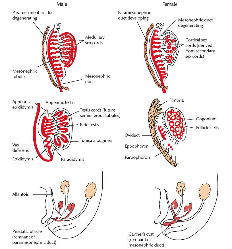
Figure 1.11 Male and female gonadal development.
The male and female genital systems are virtually identical through the seventh week. In the male, SRY protein produced by the pre-Sertoli cells causes the medullary sex cords to develop into presumptive seminiferous tubules and rete testis tubules and causes the cortical sex cords to regress. Anti-Mullerian hormone produced by the Sertoli cells then causes the paramesonephric ducts to regress and Leydig cells also develop, which in turn produce testosterone, the hormone that stimulates development of the male genital duct system, including the vas deferens and the presumptive efferent ductules.Reproduced from Robert Wilkins, Simon Cross, Ian Megson, and David Meredith, Reproduction and development, Oxford Handbook of Medical Sciences, 2011, with permission from Oxford University Press.
Later on, fetal extracellular fluid (ECF) is passed through the fetal skin and umbilical cord.
More on the topic Embryology:
- Chapter 4 Embryology and Anatomy
- REFERENCES
- Diabetes mellitus
- Thrombophilia and early pregnancy loss
- Incidence and importance
- On the Status of the Human Embryo
- References
- REFERENCES
- REFERENCES
- Contents