Pathology
Response to tissue injury
Tissue injury is associated with reversible and irreversible changes of the cell membrane. Potassium is transferred out of the cell and sodium is transferred in accompanied by water.
This leads to cellular oedema and a reduction in protein synthesis. There is a switch to anaerobic metabolism. Glycogen is used for energy resulting in lactate production and a drop in pH.Intracellular enzymes including lactate dehydrogenase, troponins, and creatinine phosphokinase become activated in association with mitochondrial damage. Lysosomal rupture results in release of lysosomal enzymes and autolysis followed by nuclear death.
Tissue growth and differentiation
Growth and differentiation aim to maintain the normal structure of a particular tissue. In tissues with continuous cell loss (blood, skin, mucosa), lost cells are continuously replaced. Stem cells frequently differentiate into a mature form during this process. In the skin, as superficial keratinized cells are shed, basal cells proliferate to replace them. The newly produced basal cells differentiate into squamous cells. When the cell turnover rate is normal,
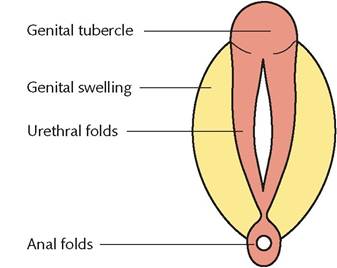
Figure 1.12 Indifferent stages of the external genitalia, approximately 6 weeks.
Reproduced from Robert Wilkins, Simon Cross, Ian Megson, and David Meredith, Reproduction and development, Oxford Handbook of Medical Sciences, 2011, with permission from Oxford University Press.
the skin appears histologically normal but if the rate is greatly increased, cells do not fully mature, and abnormalities are seen both on physical examination and histologically. The rate of cell proliferation is determined by the cell cycle and controlled by a variety of growth factors and receptors, and regulated by growth control genes.
Many of the cellular proto-oncogenes encode for growth factors for receptors.Placental pathology
The placental involvement in the pathogenesis of pre-eclampsia and particularly the link between pre-eclampsia and reduced placental perfusion has long been recognized (13).
The main defect seems to be related to endovascular trophoblast invasion. In pre-eclampsia, myometrial arteries fail to adapt to physiological change. Trophoblast invasion is impaired. Acute atherosis is common. Blood flow into the intervillous space is therefore decreased in pre-eclampsia.
Fetal growth restriction is associated with reduced uteroplacental blood flow, which in turn can be secondary to defective placentation. Microbiology
Bacteriology
Bacteria are single-cell prokaryotic microorganisms. They have a single chromosome that is not enclosed in a nuclear membrane. They can have four shapes: cocci (spheres), bacilli (rods), spirilla (spirals), and vibrios (comma-shaped). Genetic information is encoded in the cell DNA. There are two types: chromosomal DNA and extrachromosomal DNA.
Bacterial metabolism is a balance between anabolic and catabolic functions. Their ability to utilize carbohydrates and convert them to glucose as well as oxygen requirement is used to characterize bacteria:
• Obligate anaerobes: oxygen is toxic.
• Aerotolerant anaerobes: anaerobic metabolism, but tolerant to the presence of oxygen.
• Facultative anaerobes: can grow in both anaerobic as well as aerobic conditions.
• Obligate aerobes: require oxygen to grow.
• Microaerophilic organisms: require low oxygen levels only; high levels may be inhibitory.
Bacteria can be Gram positive or Gram negative (Figure 1.16). Some bacteria can suspend growth and metabolism in adverse conditions and form resistant spores. Optimum temperature range is between 20°C and 40°C.
Bacteria have four phases of growth:
1. Initial lag phase
2. Exponential growth phase
3. Static phase
4.
Death phase.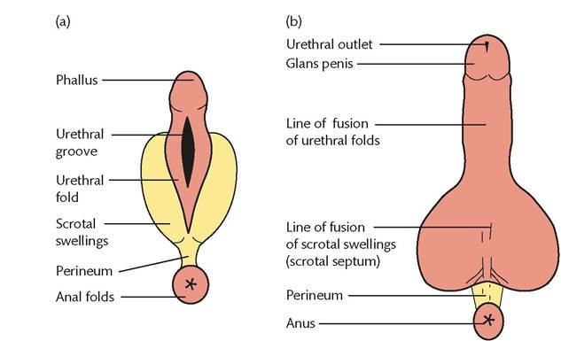
Figure 1.13 Development of the external genitalia in the male at (a) 10 weeks and (b) in the newborn. Genital tubercle extends rapidly to form the phallus, later the penis. Urethral folds close the urogenital sinus and genital swellings become scrotal swellings.
Reproduced from Robert Wilkins, Simon Cross, Ian Megson, and David Meredith, Reproduction and development, Oxford Handbook of Medical Sciences, 2011, with permission from Oxford University Press.
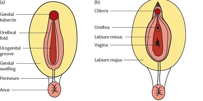
Figure 1.14 Development of the external genitalia in the female at (a) 5 months and (b) in the newborn. Genital tubercle elongates slightly, forming the clitoris. Urethral folds become labia minora and the urogenital sinus remains open. Genital swellings form the major labia.
Reproduced from Robert Wilkins, Simon Cross, Ian Megson, and David Meredith, Reproduction and development, Oxford Handbook of Medical Sciences, 2011, with permission from Oxford University Press.
Diagnosis of bacterial infection: bacteria are visible under direct microscopy. On a culture they are grown on solid agar or in liquid media and form visible colonies.
Control of infection
Antibiotics, also known as antibacterials, are used in the treatment and prevention of bacterial infections. They act by killing bacteria or inhibiting their growth. Different types of antibiotics have differences in chemical structure, mode of action, or spectrum of effect. Broad-spectrum antibiotics target a wide range of bacteria whereas narrow-spectrum ones target specific types. Antibiotics with bactericidal activities act on the bacterial cell wall (beta-l actam antibiotics: penicillins and cephalosporins as well as vancomycin and teicoplanin) or the cell membrane (polymyxins). Other bactericidal antibiotics target essential bacterial enzymes (rifamycins, lipiarmycin, quinolones, and sulphonamides).
Bacteriostatic antibiotics inhibit protein synthesis (macrolides, lincosamides, and tetracyclines).Sterilization is a process of eradication of microorganisms including the spores.
Disinfection is the removal of all actively dividing organisms. Components of disinfection include cleaning, heat, and chemicals.
Virology
A virus contains a nucleic acid (DNA or RNA) and is surrounded by a protein coat. There is no cytoplasm. Viruses must enter the host cell by endocytosis for their own replication. The virus first adheres to the host cell by binding to a specific receptor molecule.
A viral infection (Table 1.1) (14, p. 152) can cause cell death but can also cause a latent infection remaining in the host cell for many years in a dormant state.
Parasitology
Parasites are unicellular eukaryotic organisms. They can reproduce by simple asexual binary fission or a complex sexual cycle. The protozoa are larger than bacteria.
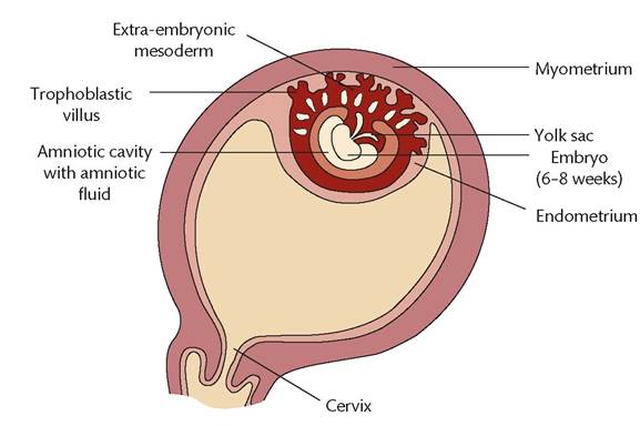
Figure 1.15 The placenta in relation to adjacent structures during early pregnancy.
Reproduced from Robert Wilkins, Simon Cross, Ian Megson, and David Meredith, Reproduction and development, Oxford Handbook of Medical Sciences, 2011, with permission from Oxford University Press.
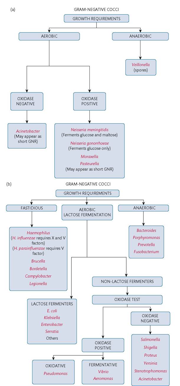
Figure 1.16 (a) Identification of Gram-negative cocci. (b) Identification of Gram-negative rods. (c) Identification of Gram-positive cocci. (d) Identification of Gram-positive rods. GNR, Gram-negative rods.
Reproduced from M. Estee Torok, Ed Moran, and Fiona Cooke, Bacteria, Oxford Handbook of infectious Diseases and Microbiology, 2016, with permission from Oxford University Press.
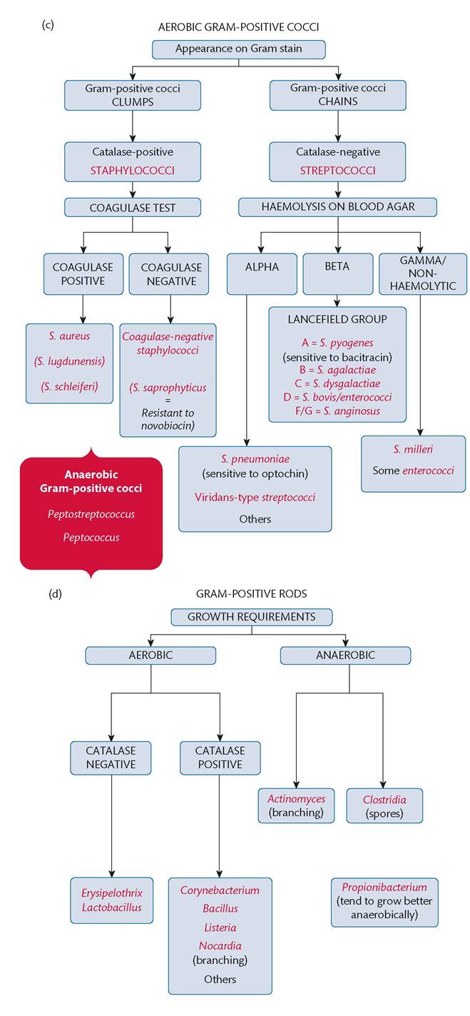
Table 1.1 Diagnosis of viral infection
| Technique | I Advantages | I Disadvantages |
| Electron microscopy | Can see if any virus present | Expensive, time-consuming, requirement for complex equipment |
| Tissue culture | Virus cultures can be made available for further analysis | Slow, expensive |
| Enzyme immunoassay | Quick, automated, immunoglobulin M assays can diagnose recent infection | Not appropriate for all viruses |
| Fluorescence microscopy | Mainly for respiratory infections | Expensive |
| Polymerase chain reaction | Quick, highly sensitive, can be automated | Expensive DNA extraction can be difficult |
Intestinal protozoa are common in environments with poor hygiene.
Entamoeba histolytica is an intestinal amoeba and causes amoebic dysentery. It spreads via the portal veins and may cause amoebic liver abscess. Giardia lamblia is a binucleate flagellate protozoan. It causes chronic diarrhoea. Cryptosporidium parvum spreads via contaminated drinking water.Malaria is caused by four types of Plasmodium: P. falciparum, P. vivax, P. ovale, and P malariae. The infection is spread by the bite of female Anopheles mosquitos and usually occurs in tropical and subtropical areas.
Trypanosoma is spread by the bite of an infected tsetse fly and is prevalent in tropical Africa. It can enter red cells, the nervous system, and the reticuloendothelial system and can also affect the myocardium. Clinical symptoms and signs include fever, drowsiness, coma, and hepatosplenomegaly.
Toxoplasma gondii is the cause of toxoplasmosis. The definitive host of the trophozoites is the cat. Reproduction takes place in its gastrointestinal tract. Humans can become infected either by handling soil contaminated by cat faeces or ingesting infected undercooked meat.
Trichomonas vaginalis is a protozoan that can cause vaginal and urethral infections. It is transmitted sexually and can cause vulvovaginal irritation with a yellow or green, frothy, ‘fishy'-smelling discharge.
Mycology
Fungi are eukaryotic organisms, commonly multicellular. The optimal growth temperature for the majority of fungi is between 25°C and 35°C. They are predominantly aerobic but many yeasts can produce alcohol by fermentation as an end product of anaerobic metabolism. They mainly reproduce by the production of asexual spores.
The main groups of pathogenic fungi are the following:
• Moulds: these form powdery colonies on culture due to the presence of abundant spores. Dermatophytes responsible for skin, hair, and nail infections belong to this group.
• True yeasts: unicellular, round or oval fungi. Reproduction is by budding from the parent cell. They appear as characteristically creamy colonies. A major pathogen in this group is Cryptococcus neoformans.
• Yeast-like fungi: these appear as round or oval cells and reproduce by budding. The major pathogen in this group is Candida, which causes vaginal candidiasis.
• Dimorphic fungi: Histoplasma capsulatum is a common member of this group. Infection is usually asymptomatic but may cause lung calcifications.