Immunology
Adaptive immunity
There are four fundamental features of adaptive immunity:
1. Memory
2. Specificity
3. Diversity
4. Tolerance to self.
Memory: the first contact with an infectious agent imparts the memory and then subsequent infection is repelled (i.e.
chickenpox). The primary response against a specific antigen occurs on first contact, is slower, and is less vigorous. Future responses are rapid and more efficient.Specificity: the adaptive immune system is specific to particular pathogens. Contact with one pathogen does not provide protection against other pathogens.
Diversity: this feature adds to the ability to combat different infections. Diversity is partly genetically encoded, but is also the result of recombination between gene segments (15) (Figure 1.17).
Tolerance: tolerance to self-antigens is established in early life. Circulating components, which reach the developing lymphoid system in the perinatal period, induce a permanent self-tolerance.
Cellular elements of adaptive immunity
T cells: these are lymphocytes derived from bone marrow stem cells which develop and differentiate in the thymus before travelling down to peripheral lymphoid tissue. There are three main types of T cells: T-helper, suppressor, and cytotoxic cells.
B cells: these are responsible for antibody production. B cells originate from stem cells in the bone marrow and develop and differentiate there.
Null cells: these lymphocytes express neither T- nor B-cell surface markers. However, they do express a mixture of lymphocyte and macrophage surface markers. They bind via the Fc receptor and kill the target cells. Hence, they are also called killer cells.
Antigen-presenting cells (APCs): antigen presentation is the process by which cells express molecules recognizable by T cells. These APCs take up antigen and process and modify it into an immunogenic form before presenting it along with major histocompatibility complex molecules to T cells.
Immunoglobulins, complement, and cytokines are the three principal humoral elements of adaptive immunity.
Innate immunity
Cellular elements of innate immunity are phagocytes and natural killer (NK) cells. The soluble elements are complement, acute phase proteins, and interferon.
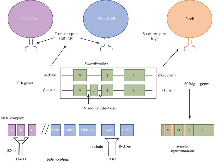
Figure 1.17 Generation of diversity in the immune system. Both T-cell receptors (TCRs) and B-cell receptors (BCRs) are generated through recombination. For immunoglobulin (Ig)-H chains there are 65, 27, and 8, variable (V), diversity (D), and joining (J) genes encoded in the germ line respectively. Somatic hypermutation occurs only in B cells. The major histocompatibility complex (MHC) complex is highly polymorphic—the class II genes actually lie upstream of the class I genes on chromosome 6. DRA encodes the α chain which is conserved and pairs with polymorphic β chains from the DRB1 locus.
Reproduced from Paul Klenerman, Adaptive immunity, in: Oxford Textbook of Medicine 5e (eds. David Warrell, John Firth, Timothy Cox). Oxford University Press 2010, with permission from Oxford University Press.
Phagocytes are immune cells that phagocytose pathogens by engulfing, destroying, and processing before presenting to cells of the adaptive immunity. They are derived from bone marrow stem cells.
NK cells: these leucocytes destroy compromised host cells. They recognize cell surface changes on tumour cells or virus-infected cells. They engage and kill these cells.
Complement: this is an essential component of the innate immune system and consists of more than 30 interacting proteins and receptors. It can be activated by antigen-antibody interactions or by microorganisms. There are various mechanisms of function: some adhere to pathogens and promote phagocytosis, some attract phagocytes to the site of the reaction (chemotaxis), and some lyse the cell membranes of bacteria.
Pathways of complement activation include the classical pathway, the alternative pathway, the lectin pathway, and the terminal pathway (Figure 1.18).
Regulation of the immune system
Antigens taken up by APCs are presented to T-helper cells. The APCs release cytokines, which stimulate T cells and interleukin-2 receptor expression on the cells. T- helper cells help B cells to produce antibodies, which can neutralize the antigens. The antibodies can also promote the ingestion of antigens by macrophages. Cells infected by viruses can also be attacked by cytotoxic T cells, macrophages, or NK cells. The level of response is regulated by antibodies, suppressor T cells, APCs, and helper T cells. The primary purpose of such responses is to minimize damage caused by pathogens, eliminate pathogens, and establish future memory.
Pregnancy and immunity
The fetus contains antigens of both maternal and paternal origin and yet is not rejected by the mother. It is now clear that the fetus is antigenically mature from an early stage and that immunocompetence begins to develop in the first trimester of pregnancy. The uterus is not immunologically privileged. It has an extensive lymphatic and vascular network especially prominent during pregnancy.
There is ample evidence for a decrease in the maternal immune response in pregnancy. However, pregnant women are still sufficiently immunocompetent to remain healthy. This effect occurs from the time of implantation when the endometrium decidualizes. Only two types of fetoplacental tissue come into direct contact with maternal tissues: the villous trophoblast and EVT. There are
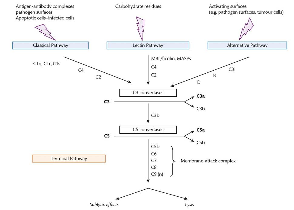
Figure 1.18 Simplified overview of the complement system showing the three main activation pathways and the terminal pathway culminating in the formation of C5b-9.
Reproduced from Marina Botto and Mark J. Walport, The complement system, in: Oxford Textbook of Medicine 5e (eds. David Warrell, John Firth, Timothy Cox). Oxford University Press 2010, with permission from Oxford University Press.
effectively no systemic maternal T- and B-cell responses to trophoblast cells in humans. The villous trophoblast, bathed by maternal blood, seems to be immunologically inert. It never expresses human leucocyte antigen (HLA) class I or class II molecules. EVT, which is directly in contact with maternal decidual tissues, does not express the major T-cell ligands, HLA-A or HLA-B, but does express the HLA class I trophoblast-specific HLA-G, which is strongly immunosuppressive.
A population of NK-derived, CD56 granulated lymphocytes is found in the first trimester. They release transforming growth factor beta-2, which also has immunosuppressive activity.
More on the topic Immunology:
- HYPERSENSITIVITY DISORDERS
- RENAL VEIN THROMBOSIS
- Shilifoshi, San foqi, and Malayu
- Macrovascular Complications of Diabetes Mellitus
- PASSIVE IMMUNIZATION
- Conclusion
- Prevalence and risk factors
- Pathogenesis
- phylum Nematoda
- Corynebacterium bovis Infection: Coryneform Hyperkeratosis; Scaly Skin Disease