Trauma in Pregnancy
Victor J. Hassid and Miren A. Schinco
Trauma is the primary cause of death in women of reproductive age and is the leading nonobstetric cause of both fetal and maternal death during pregnancy (1-4).
The potential for pregnancy must be considered in any girl or woman between the ages of 10 and 50 years. Accidental injury is estimated to occur in 6% to 7% of all pregnancies, and its incidence increases as pregnancy progresses until, by the end of the third trimester, minor trauma occurs more frequently than at any other time during female adulthood (1,2,5-8).The majority of blunt trauma during pregnancy results from motor vehicle crashes, with the remainder being relatively evenly distributed between falls and assaults (3,4,9-13). Some authors report that up to 11% of women are victims of physical abuse during pregnancy (14-17) and up to 31.5% of pregnant women admitted following trauma are victims of intentional injury (11). During the third trimester, an altered center of gravity (caused by the enlarged uterus) combined with pelvic ligamentous laxity produces a degree of gait instability that makes the pregnant woman particularly susceptible to injury by falls (1,9). This represents the second most common mechanism of injury during pregnancy (13).
In a recent review of over 77,000 women of childbearing age in the American College of Surgeon’s National Trauma Data Bank, pregnant patients were significantly younger, more likely to be underinsured, and of African American or Hispanic descent than their nonpregnant counterparts. While they were less likely to have used alcohol or illicit drugs, still 13% had been drinking and 20% had used drugs prior to their trauma. Although pregnant trauma patients were more likely to use seat belts, still only two thirds were restrained. After admission for a trauma, 5.1% of patients went on to delivery, three quarters within 24 hours and by cesarean section.
The foundation of trauma management in pregnancy is that “the best chance for fetal survival is to assure maternal survival” (5). The physician caring for a pregnant trauma patient should always remember that there are two patients. Initial treatment priorities for an injured pregnant patient remain the same as for the nonpregnant patient. The use of imaging studies should not be withheld because of pregnancy. Because the anatomic and physiologic alterations of pregnancy can alter the gravid woman’s response to injury, an understanding of these changes is critical when approaching trauma management. Early communication between the trauma surgeon and the obstetrician is very important in order to optimize the management of the pregnant trauma patient.
OUTCOMES
Regardless of severity, trauma during pregnancy has been associated with spontaneous abortion, premature labor, uterine injury, placental abruption, maternal death, and low birth weight fetal loss. When comparing traumatized gravid women with nontrauma controls, for any injury, both maternal and fetal outcomes are worse, with progressively worse outcomes with higher injury severity score (ISS). Injuries associated with the highest maternal mortality are thoracoabdominal and pelvic followed by brain injuries. Women who sustain trauma, but do not deliver at that time, continue with significant risk of increased morbidity and mortality throughout their pregnancy and therefore require close monitoring (13).
Maternal predictors of fetal death consist of maternal factors (ISS >15, severe head injury, pelvic fractures, shock, hypoxemia, and absent fetal heart tones) and mechanisms of injury (auto-pedestrian collisions, maternal ejection, motorcycle collision, and lack of restraints) (18-21). Infants under 28 weeks’ gestational age appear to have the highest potential for complications, possibly due to their intolerance of maternal stress (13).
ANATOMIC AND PHYSIOLOGIC ALTERATIONS OF PREGNANCY
Anatomic Changes
Until approximately the 12th gestational week, the uterus remains in the pelvis, and by 20 weeks and 34 to 36 weeks, it reaches the umbilicus and the costal margin, respectively.
During the last 2 weeks of gestation, the fundus frequently descends as the fetal head engages the pelvis. The progressive enlargement of the uterus causes the bowel to be displaced cephalad and therefore keeps it relatively protected from blunt abdominal trauma, whereas the uterus and its contents become more vulnerable (Fig. 4.1).During the second trimester, the uterus enlarges beyond its protected intrapelvic location, but the fetus remains mobile and protected by a generous amount of amniotic fluid; the amniotic fluid can be a source of embolism and disseminated intravascular coagulation following trauma if it gains access to the intravascular space.
By the third trimester, secondary to the location of the fetal head within the pelvis, pelvic fractures may result in fetal skull fracture or significant intracranial injury. Unlike the elastic myometrium, the placenta has little elasticity, which predisposes it to shear forces at the uteroplacental interface and placental abruption.
While interpreting pelvic x-rays, it is important to remember that the pubic symphysis widens to 4 to 8 mm and the sacroiliac joint spaces increase by the 7th month of gestation.
Cardiovascular System
Maternal cardiac output begins to increase during the first 10 weeks of pregnancy, reaching a peak of 30% to 50% above nonpregnant levels by the latter part of the second trimester (22). After the 10th week of pregnancy, the cardiac output can be increased by 1.0 to 1.5 L per minute, due to the increase in plasma volume and decrease in vascular resistance of the uterus and placenta, which during the third trimester of pregnancy receive 20% of the patient’s cardiac output. During the same period, the cardiac output may be transiently lowered by aortocaval compression by the gravid uterus, resulting in decreased placental perfusion. This phenomenon, known as the supine hypotension syndrome, may be observed clinically in otherwise normal pregnant patients (22,23). It is very important to remember while managing a pregnant trauma patient that changing the maternal position from the supine to the left lateral decubitus position may increase cardiac output by as much as 25% at term (23).
The average maternal heart rate increases 20% to 30% during pregnancy such that by term, the normal maternal pulse rate is 80 to 95 beats per minute, making borderline tachycardia difficult to evaluate (2,9,14,23). Mean arterial blood pressure gradually declines during the first two trimesters of pregnancy, reaching its nadir by approximately 28 weeks’ gestation (22).
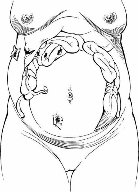
FIGURE 4.1 Compartmentalization of intestines during pregnancy and sites of gunshot wounds above and below the umbilicus.
Systolic arterial pressure decreases by 0 to 15 mm Hg, whereas diastolic pressure declines by 10 to 20 mm Hg, creating a widened pulse pressure. These changes are the result of diminishing peripheral vascular resistance and should not be mistaken as evidence of hypovolemia during the first two trimesters of pregnancy. During the third trimester, blood pressure gradually increases, returning to pre-pregnancy levels near term (2,7,9,22,23). Hypertension, systolic or diastolic, is never expected during pregnancy and, if present, may either be a response to pain, anxiety, or injury or be the result of a direct complication of pregnancy such as pregnancy-induced hypertension (9).
Associated with the underlying decreased peripheral vascular resistance of the first two trimesters of pregnancy is a paradoxical response to stimuli that would normally cause vasoconstriction. This altered response causes the skin to be warm and dry, instead of cool and clammy which would be expected during hypovolemic shock (9). In addition, central venous pressure, normally approximately 9cm ^O in the nonpregnant patient, gradually decreases throughout pregnancy until it reaches 4 to 6 cm ^O during the third trimester (4,7,14,24). Venous hypertension in the lower extremities is present during the third trimester.
Total maternal blood volume increases by as much as 50% at 34 weeks’ gestation, improving maternal response to hemorrhage.
A smaller increase in red blood cell (RBC) volume occurs, resulting in a decreased hematocrit (physiologic anemia of pregnancy) (1,22). The white blood cell (WBC) count increases, resulting in counts as high as 15K and 25K∕mm3 during pregnancy and labor, respectively (1,5,9). Levels of serum fibrinogen and other clotting factors are mildly elevated, leading to a hypercoagulable state (14,22). During periods of stress, the mother maintains homeostasis at the expense of the fetus. Acute maternal hemorrhage or maternal hypoxia induces uterine artery vasoconstriction, which can reduce uterine perfusion by 10% to 20% before clinical evidence of maternal hypovolemia occurs (1,4,5,9). Consequently, 30% to 35% of maternal blood volume may be lost before clinical signs of hypovolemia develop (1,6,7,9,14,24). This, combined with the sensitivity of the placental vasculature to catecholamines, places the fetus at risk during maternal hemorrhage that may, upon clinical examination of the mother, appear minimally significant. Early and adequate maternal volume replacement and thorough fetal evaluation, including fetal monitoring, are therefore critical in the management of the gravid trauma patient.Electrocardiographic (ECG) changes in late pregnancy are not specific and reflect the leftward shift of the cardiac axis, by approximately 15 degrees, due to diaphragm elevation. The T waves may be flattened and inverted in lead III, and the Q waves may appear in leads III and aVF. Supraventricular ectopic beats may also be seen (9,14).
Respiratory System
Minute ventilation increases by 40% to 50% over control values at term due to increases in tidal volume and respiratory rate (9,14,22,25). The metabolic effect of this change is a partially compensated respiratory alkalosis, with arterial blood gases showing a decrease in PCO2 to approximately 30 to 34 mm Hg and a decrease in serum bicarbonate to 18 to 22 mEq∕L. The net effect is to leave the pregnant woman with a decreased buffering capacity after trauma (1,9,22).
A PaCO2 of 35 to 40 mm Hg may indicate impending respiratory failure during pregnancy. Maternal arterial PO2 increases by 10 mm Hg, but oxygen consumption increases by as much as 20% by term (9,14,25). Coupled with fetal oxygen demand and fetal sensitivity to hypoxia, these alterations place the pregnant trauma patient and her fetus at risk for hypoxic insult. Supplemental oxygen therefore becomes a priority in the pregnant trauma patient (9,25). Anatomic alterations in the thoracic cavity appear to account for the decreased residual volume, associated with diaphragmatic elevation, increased lung markings, and prominence of the pulmonary vessels noticed on chest x-ray. These should be kept in mind when considering chest tube placement in a pregnant trauma patient.Gastrointestinal System
The major physiologic alterations of the gastrointestinal system are those of increasing compartmentalization of the small intestine into the upper abdomen and a progesterone-induced decrease in gastrointestinal motility. The gravid uterus may protect the abdominal viscera from injury to the lower abdomen, but penetrating wounds to the upper abdomen may injure many loops of tightly crowded small intestine (Fig. 4.1). In addition, stretching of the abdominal wall alters the normal response to peritoneal irritation, at times masking significant intra-abdominal organ injury. Decreased gastric emptying increases the possibility of aspiration during trauma and intubation, therefore early gastric tube decompression is important in order to prevent aspiration of gastric contents (1,7,9,22,24,25). Position of the patient’s spleen and liver is essentially unchanged by pregnancy.
Urinary System
Progesterone-induced smooth muscle relaxation of the ureters and mechanical compression by the gravid uterus cause dilatation of the ureters and renal pel- vises, which may appear as hydronephrosis on radiographic studies (14,22). In addition, ureteropelvic dilatation predisposes the urinary collecting system to stasis and subsequent infection, particularly after catheterization (14,22). Anatomically, the urinary bladder is displaced anteriorly and superiorly by the enlarging uterus, making it increasingly vulnerable to injury after the first trimester (1,2,8,22). Furthermore, the glomerular filtration rate and the renal plasma blood flow increase during pregnancy and the levels of creatinine and serum urea nitrogen drop to approximately one half of normal pre-pregnancy levels.
Endocrine System
The pituitary gland increases in size and weight by 30% to 50% during pregnancy, and any significant drop in blood pressure can result in necrosis of the anterior pituitary gland and subsequent pituitary insufficiency.
Neurologic System
One of the complications of late pregnancy is eclampsia, which can present with the same signs and symptoms as a head injury. It should always be considered in case of seizures associated with hypertension, hyperreflexia, proteinuria, and peripheral edema. It may be necessary to obtain expert neurologic consultation in these cases in order to differentiate between eclampsia and other causes of seizures.
Table 4.1 outlines the physiologic, anatomic, and laboratory changes associated with pregnancy.
Patterns of maternal and fetal injury
Blunt Abdominal Trauma
Following blunt abdominal trauma during the latter part of pregnancy, the gravid uterus is subject to direct injury, as well as to the shearing forces resulting from sudden deceleration. Most fetal morbidity is a result of catastrophic maternal trauma; however, some serious complications, including preterm delivery, abruptio placentae, fetal injury, fetal death, and massive fetomaternal hemorrhage (FMH), have occurred after seemingly minor injuries (5,11,26,27).
The abdominal wall, uterine myometrium, and amniotic fluid act as buffers to direct fetal injury from blunt trauma. Still, fetal injuries can occur when the abdominal wall strikes the dashboard or steering wheel, or in case, the pregnant patient is struck by a blunt instrument. Indirect fetal injury may take place secondary to rapid deceleration, contrecoup effect, or a shearing force leading to placental abruption.
TABLE
4.1
Physiologic and Anatomic Changes in Pregnancy
| System | Physiologic Change | Effect |
| Cardiovascular | ↑ Cardiac output | Tachycardia |
| ↑ Blood volume | ↑ Maternal tolerance to hemorrhage | |
| T Peripheral vascular | T Blood pressure; ↑ skin | |
| resistance | temperature | |
| T Central venous pressure | 4-6 cm H2O during third trimester | |
| Aortocaval compression | Supine hypotension | |
| T Uterine perfusion in response to hemorrhage | ↑ Fetal risk during hemorrhage | |
| Pulmonary | ↑ Minute ventilation | Partially compensated respiratory alkalosis; T tolerance to acidosis |
| ↑ Oxygen consumption ↑ Fetal oxygen demand | T Tolerance to hypoxia | |
| Elevation of diaphragm 4 cm by | Thoracostomies performed one | |
| term | to two interspaces higher than usual | |
| Gastrointestinal | T Motility; T gastric emptying compartmentalization | ↑ Risk of aspiration ↑ Risk of injury with upper abdominal trauma T Risk with lower abdominal trauma |
| Genitourinary | T Motility of collecting system; | Physiologic abnormality of |
| T bladder emptying | radiographic studies ↑ Risk of infection with catheterization | |
| Displacement of bladder | ↑ Risk of injury | |
| ↑ Uterine size and blood | ↑ Risk of hemorrhage from | |
| flow | abdominal wounds | |
| Hematologic | ↑ Plasma volume without proportional ↑ in red cell mass | Dilutional anemia |
| Leukocytosis | Normal WBC count can reach 18,000/mm3 in second and third trimester | |
| ↑ Factors VII, VIII, IX, and X and fibrinogen | Hypercoagulable state |
Note: ↑ Increased; T decreased. WBC, white blood cell.
Abruptio placentae is a significant cause of fetal loss in both catastrophic and noncatastrophic trauma. While the exact mechanism of traumatic abruption is not known, the suggested mechanism is based on the fundamental differences in tissue characteristics between the relatively elastic myometrium of the uterus and the relatively inelastic tissue of the placenta. When an external deforming force is applied to the abdomen, shearing of the uteroplacental interface occurs. Shearing is further aggravated by the increased intrauterine pressure that results from impact (10,15). Signs and symptoms suggesting abruption include vaginal bleeding, uterine tenderness or contractions, fetal heart rate abnormalities, and fetal death. Although the presence of these symptoms is significant, the absence of symptoms following trauma does not exclude the possibility of placental abruption (3,27-29). Most cases of significant abruption can be identified by clinical signs or electronic fetal monitoring within 4 to 6 hours of the traumatic event (15,23,30), however, even in minor abdominal trauma, significant placental abruption can occur without significant symptoms. This supports the i mportance of fetal monitoring after abdominal trauma (20). Cases of abruptio placentae have been reported to occur up to 5 days following severe trauma (29,31).
Uterine rupture may also result from blunt trauma. Uterine rupture complicates approximately 0.6% of traumatic events during pregnancy and tends to occur only with major blunt abdominal trauma (15,23). The presentation of uterine rupture ranges from subtle findings such as uterine tenderness and worrisome fetal heart rate patterns, without changes in maternal vital signs, to rapid onset of maternal hypovolemic shock associated with fetal and maternal death (15). Fetal mortality rate approaches 100%; maternal mortality is usually due to concurrent injury (23).
Amniotic fluid embolism is a rare complication of pregnancy characterized by poor response to treatment and high mortality. The incidence is between 1 in 8,000 and 1 in 80,000 live births with mortality ranging from 61% to 86%. While it most frequently occurs in the peripartum period, it has also been described after blunt abdominal trauma. It typically presents with sudden onset of hypoxia, altered mental status, hemody namic compromise, and disseminated intravascular coagulation. The diagnosis remains largely a clinical one. Management is mainly nonspecific supportive therapy aimed at cardiopulmonary resuscitation, correction of coagulopathy, and treatment of hemorrhage. If delivery has not occurred, emergent cesarean section prevents further hypoxic insult to the fetus and facilitates treatment of the mother (32).
Premature uterine contractions are another common sequela of maternal trauma (1,3,30-31,33-36). Studies have shown that up to two thirds of traumatized gravidas experience frequent contractions during the initial 4 hours of monitoring (3,30,34). Postulated etiologies include placental abruption, uterine contusion, membrane ischemia, and membrane rupture (1). The use of tocolytics to halt premature contractions associated with trauma is controversial. Although some authors report successful cessation of preterm contractions with these agents (36,37), others discourage their use, believing that regular uterine activity after trauma will spontaneously subside during observation. In those cases in which contractions persist, placental abruption must be considered until proven otherwise (14,34). Although the mean abdominal abbreviated injury score (aAIS), a direct indicator of injury severity of intra-abdominal structures, is usually higher among patients who sustained acute termination of pregnancy and/or fetal loss, in some studies, a high aAIS (≥3) did not independently predict these complications (38).
Direct fetal injury complicates the effectiveness of seat belts during pregnancy (50).
Penetrating Abdominal Trauma
Gunshot and stab wounds are the most frequently encountered penetrating injuries (14,22). As with blunt trauma, pregnancy often changes the usual manifestations of penetrating abdominal trauma because the gravid uterus displaces other abdominal organs cephalad. When the route of entry is below the fundus, the gravid uterus will often protect maternal viscera by absorbing the force of the shell or instrument. In contrast, upper abdominal injuries may result in increased injury to bowel compressed into the upper abdomen (14,22).
Gunshot wounds to the abdomen are associated with a high rate of internal organ injury. When a bullet, particularly one of low velocity such as that commonly found in civilian injuries, strikes the pregnant uterus, much of its energy is dissipated in the dense uterine musculature, diminishing its velocity and ability to penetrate other maternal abdominal organs. Gunshot wounds to the pregnant abdomen therefore are associated with low maternal rates of morbidity and mortality (4,24,53). Fetal outcome, however, is generally poor, with reported rates of fetal injury varying from 59% to 78% and perinatal mortality ranging from 41% to 71% (1,22,24,39,53). Fetal mortality is caused by both prematurity and direct fetal injury (24). Although direct fetal injury portends a poor prognosis, cases have been reported in which a fetus having sustained a nonfatal injury later was successfully delivered alive (7,24,54). Because gunshot wounds to the abdomen have a high frequency of internal organ injury, surgical exploration remains the standard approach to this type of injuries (4,15,16,22).
Stab wounds to the abdomen are associated with a lower mortality rate than are gunshot wounds. As with gunshot wounds, the gravid uterus may protect the victim of a lower abdominal stab wound, whereas wounds to the upper abdomen carry a higher incidence of visceral injury (4,7,15,22). The trend toward conservative management of abdominal stab wounds extends to the pregnant patient. Clinical considerations influencing management include gestational age, condition of the mother and the fetus, and location of the wound in the abdomen. Management options include immediate surgical exploration, in case of diffuse abdominal tenderness or presence of peritonitis, or close observation and use of adjunctive imaging studies, which may provide information regarding potentially injured viscera and penetration or not of the abdominal fascia.
EVALUATION AND MANAGEMENT
Initial evaluation and resuscitative efforts should be directed toward the mother, because the most common cause of traumatic fetal death is maternal death. Once maternal airway, respiratory, and hemodynamic stability are ensured, attention must be immediately directed to assessment and assurance of fetal health.
Prehospital Management
Injured pregnant women, particularly those in the latter half of pregnancy, should be transported to an appropriately designated trauma center with facilities for adequate maternal and specialized neonatal care when possible (4,9). During the second and third trimesters, transport should be performed with the patient in the left lateral decubitus position or the spine must be immobilized, with the backboard tilted 15 degrees to the left (1,7,9,14). Listening for fetal heart tones may be performed en route but should not delay transport (7,14). The pneumatic antishock garment (PASG) may be used to control bleeding from pelvic or lower extremity fractures, but its use should not interfere with rapid reestablishment of intravascular volume by intravenous fluid infusion. If it is used during pregnancy, only the lower or leg compartments should be inflated. Inflation of the abdominal compartment is contraindicated in any patient with a potentially salvageable gestation because the PASG has a deleterious effect on uteroplacental blood flow, compromises maternal venous return, and has the potential to cause direct fetal injury (1,4,7,9,22,23).
Both in the prehospital setting and in the emergency department, oxygen therapy provides special benefit for the fetus. Although the maternal arterial blood is normally well saturated with oxygen when the mother is breathing room air, the oxygen hemoglobin dissociation curve of fetal blood is shifted such that increasing alveolar oxygen tension continues to improve fetal saturation and thereby fetal outcome (7,14). Maternal hemorrhage can reduce uterine blood flow 10% to 20% before signs of maternal hypovolemia appear (1,4,5,9). Therefore, early intravenous access and volume resuscitation are important for both the mother and the fetus. Vasopressors should be avoided, because they can further decrease uterine blood flow (4,7,15). The presence of initial maternal hypotension (defined as systolic blood pressure ≤90 mm Hg) has been shown to be associated with a higher risk of pregnancy loss. Toward the third trimester, maternal intravascular volume increases up to 50%. This improves maternal tolerance of hemorrhage and delays the appearance of signs and symptoms of shock, while the uterine circulation is reduced. Once obvious maternal hypotension ensues, the fetus has already been endangered by decreased uterine arterial blood flow (55).
Evaluation of the Mother and the Fetus
Prehospital information is critical in ascertaining the mechanism of injury, vital signs, and patient condition in the field. Prenatal history is of particular importance, particularly in ascertaining the gestational age of the fetus and the presence of complicating medical conditions. Maternal perception of fetal movement may provide a very cursory indication of fetal health. The time of the last meal is more significant than in the nonpregnant patient because of the prolonged gastric emptying time associated with pregnancy (7,9).
Initial physical assessment of the mother is focused on the stability of the airway, adequacy of ventilation, effectiveness of the circulatory volume, and evaluation for occult injuries; there exist conflicting data regarding the relationship between the mother’s Glascow Coma Scale (GCS) and the fetal outcome (56). Supplemental oxygen should be administered to the mother throughout the initial resuscitation and evaluation, because minimal changes in maternal oxygenation result in appreciable changes in fetal oxygen content and reserve (4,14,25). Because of the increased potential for aspiration and importance of fetal oxygenation, endotracheal intubation should be pursued more aggressively than in the nonpregnant patient (4,15,22,25). The expanded maternal intravascular volume associated with pregnancy may delay the onset of clinical signs of shock, so fluid resuscitation should be more aggressive than in nongravid patients. Crystalloid fluid resuscitation and early type-specific blood administration are indicated to support the physiologic hypervolemia of pregnancy. Non-cross-matched type 0, Rh D-negative blood can be used if type-specific blood is not immediately available (16,23). Continuous uterine displacement, either manually or by placing the mother in the left lateral tilt position, is desirable after 20 weeks’ gestation, because of the uterine compression of the inferior vena cava. Vasopressors should be avoided until appropriate volume replacement, including blood transfusion if indicated, has been administered (4,14,15,22).
Once maternal hemodynamic stability has been established and assessment of maternal injuries has begun, the condition of the fetus should be ascertained. Fetal evaluation includes determining fetal heart rate and fetal movement, evaluating uterine size and irritability, and examining the vagina for vaginal bleeding or leakage of amniotic fluid. Ideally, fetal heart rate and maternal contractions should be continuously monitored using external tococardiography. However, when this is not available, fetal heart tones should be auscultated at frequent intervals. Fetal heart rate abnormalities including tachycardia, late decelerations, and ultimately fetal bradycardia, even in the absence of vaginal bleeding and uterine tenderness or irritability, are highly suggestive of placental abruption (4,14).
Determination of fetal age and maturity may be critical in management decisions for the fetus. The quickest means of estimating gestational age is by palpation of the fundal height. At 12 weeks’ gestation, the uterine fundus rises just above the symphysis pubis; at 20 weeks’ gestation, it reaches the level of the umbilicus; and by 36 weeks of gestation, it lies just below the xiphoid process. The age of fetal viability is generally considered to be 24 to 26 weeks’ gestation, depending, in part, on the neonatal facilities available (1,2,7). Palpation of the uterine fundus two to three finger-breadths above the umbilicus is consistent with the presence of a potentially viable fetus. Ultrasound is a more accurate tool and should be used if circumstances permit (4,14).
Asymptomatic patients with minor or insignificant abdominal trauma may be discharged with reassurance after 4 to 6 hours of fetal and maternal monitoring. Discharge instructions should include the need to return upon the development of abdominal pain, decreased fetal movement, vaginal bleeding, or leakage of fluid. Timely obstetric follow-up is advised (15,16).
Routine Laboratory Studies
Urine β-human chorionic gonadotropin testing should be performed on all female trauma patients of childbearing age who are not obviously pregnant, as approximately 3% of females admitted to a trauma center are pregnant, and 8% of these pregnancies are newly diagnosed (57,58). Routine laboratory studies in the pregnant patient with significant trauma include type and cross-match; complete blood count with platelet count; electrolytes; and coagulation panel including prothrombin time, partial thromboplastin time, fibrinogen level, and fibrin degradation products (1,9,14,16,23). The finding of a normal nonpregnant value of fibrinogen (200 to 250 mg/dL) may be consistent with early disseminated intravascular coagulopathy (14), because normal fibrinogen levels during pregnancy may be twice that in nonpregnant women. Rh D-negative patients typically will undergo K-B testing to determine the volume of FMH that has occurred (14,15,22,36,43).
Radiographic Procedures
The first trimester of intrauterine development is the time of greatest sensitivity to radiation (59). During the first 2 weeks of conception, the embryo is particularly sensitive to the lethal effects of radiation but has a low probability of sustaining teratogenic effects if implantation and continued development occur (60,61). During the period of embryonic development (2 to 8 weeks after conception), the embryo is particularly sensitive to the growth-retarding and teratogenic effects of radiation; from the 8th to the 15th week after conception, the fetal brain is undergoing rapid neuron development and is potentially vulnerable to radiation-induced mental defects. Throughout gestation, there is an increased risk of childhood malignancies, especially leukemia, in children exposed to radiation in utero (60-62). Determining the exact incidence of adverse effects, however, is difficult because of the prevalence of congenital malformations occurring in the general population and because of the other frequent biomedical and social factors that may influence pregnancy outcome (59,60). In general, fetal risk is considered negligible when the total radiation exposure is ≤5 rad and the risk of malformations is significantly increased above the control levels only at doses >10 to 15 rad (14,15,48,63). An increased risk of childhood cancers has been found at somewhat lower radiation doses (22,60,62). The dose of radiation absorbed by the fetus depends on several factors including the (a) energy and intensity of the x-rays, (b) proximity of the uterus to the anatomic area exposed, (c) extent of the area exposed, (d) patient positioning, (e) depth of the fetus, and (f) number of films acquired (64). If the uterus is outside the area radiographed, the dose of radiation to the conceptus is limited to that delivered by scatter and leakage radiation, declining rapidly farther the uterus is from the radiographed area. Pelvic shielding should be used whenever possible (9). Estimates of fetal radiation exposure from commonly used procedures are listed in Table 4.2. Routine diagnostic radiographs seldom result in significant radiation exposure to the fetus. Therefore, although unnecessary
Estimated Radiation Dose to the Uterus From Commonly Used Radiographic Examinations
| Examination | Typical Dose (mrad/ Examination)3 | Range (mrad/Examination)3 |
| Cervical spine | fluid volume, and fetal movement, but it is not as sensitive as cardiotocographic monitoring for diagnosing early and clinically significant abruptio placentae (4,23). Although small abruptions are not easily seen, large abruptions, retroplacental clots, and some fetal fractures may be identified (1,2). Fetal Monitoring Continuous fetal monitoring with a cardiotocodynamometer is recommended for all viable fetuses after 20 to 24 weeks’ gestation. The normal range for fetal heart rate is between 120 and 160 beats per minute. An abnormal heart rate, repetitive decelerations, absence of accelerations or beat-to-beat variability, or frequent uterine activity may be a sign of impending maternal and/or fetal decompensation (1,2). The duration of monitoring remains controversial. Patients at risk for an adverse fetal outcome and for placental abruption require prolonged evaluation. Several studies have shown that most complications reveal themselves within 6 hours of the traumatic event. So, patients found to be at risk should undergo continuous monitoring for 6 hours. If there is no evidence of adverse fetal outcome, they may be discharged home with an instruction to return immediately in case of any signs of placental abruption, bleeding, contraction, or change in fetal movements. Patients with evidence of contractions, requiring general anesthesia and those at significant risk for unfavorable outcomes or placental abruption, must undergo 24 hours of continuous fetal monitoring. Any evidence of fetal distress should be managed aggressively with delivery by cesarean section, if necessary (21). Peritoneal Lavage Diagnostic peritoneal lavage (DPL) has largely been supplanted by the FAST examination. However, DPL is a safe and accurate procedure in the pregnant trauma patient and can be considered in cases of blunt abdominal trauma as well as anterior abdominal and thoracoabdominal stab wounds (4,26). Open lavage, which allows direct visualization of the peritoneum at a site in the midline just above the uterine fundus, is usually recommended (4,5,7,15). Unless gross blood is obtained from the initial aspirate, a peritoneal dialysis catheter is placed above the umbilicus using the open technique, and it should be gently directed toward the pelvis and a liter of crystalloid infused through it. Fluid is returned by gravity drainage. Criteria for a positive lavage are identical to that in a nonpregnant patient (5). DPL does not assess retroperitoneal and intrauterine pathology, which may be better visualized by CT scanning in the stable patient with blunt abdominal trauma or penetrating wounds of the flank and back (4). Tetanus Prophylaxis Transplacental transfer of tetanus antitoxin prevents neonatal tetanus (65). Therefore, tetanus immunization is recommended for both postexposure and routine administration during pregnancy. Indications, dose, and timing for postexposure tetanus prophylaxis with combined tetanus and diphtheria toxoids and passive immunization with tetanus immunoglobulin are identical to those in the nongravid female (65,66). Although there is no evidence that tetanus and diphtheria toxoids are teratogenic, routine administration of diphtheria toxoids should be performed during the second or third trimester to minimize concerns over teratogenicity (67). Injury Severity Score The ISS is used frequently for injury severity assessment. Attempts to use the ISS to predict placental abruption or fetal death have had conflicting results, but the weight of evidence points to a correlation between a higher ISS and a higher risk of fetal death (53,68,69). Ejection from the vehicle, motorcycle crashes, pedestrian collisions, and maternal death do appear to be independent risk factors for fetal demise (70). Increased maternal age, loss of consciousness, and maternal pelvic fractures sustained during motor vehicle collisions contribute toward high ISS and impact fetal outcome (56). One study concluded that the only combination which could reliably exclude the possibility of fetal mortality was an ISS 20 weeks’ gestation who has these symptoms should be suspected of having eclampsia and should be treated as such, while undergoing evaluation and management for suspected head injury. Although hypertension, edema, proteinuria, and hyperactive reflexes are frequently present, they may not be prominent. When eclampsia is suspected, treatment with magnesium sulfate (MgSO4) should be initiated immediately, per protocol. Urinary output, respiratory status, and the presence of patellar reflexes should be monitored during MgSO4 infusion. Injury to the spinal cord is not uncommon either as an isolated injury or in combination with other blunt or penetrating injuries. These injuries and the accompanying neurologic deficit or neurogenic shock may be difficult to diagnose in patients with an altered level of consciousness. A high index of suspicion should be maintained. Refractory hypotension unresponsive to proper patient positioning (15 degrees to the patient’s left) or aggressive fluid replacement, without an identified ongoing source of blood loss, should raise the possibility of neurogenic shock. Thermal Burns Although thermal burns do not commonly complicate pregnancy, it is estimated that between 5% and 10% of women of reproductive age admitted to burn units with significant burns are pregnant (39). Several observations have emerged from published studies of gravid patients who have sustained thermal injury (71-73). First, pregnancy does not appear to alter maternal outcome after thermal injury (14,22,71,73). Second, as might be expected, maternal and fetal survival are inversely related to the extent of the mother’s burn (71-73), with burns covering >50% of the total body surface area portending a poor prognosis for the fetus (14,39,72). Premature labor or fetal death generally occurs within the 1st week after the burn injury and is often preceded by episodes of maternal hypotension, sepsis, or respiratory failure (39,72,73). As in other forms of trauma, fetal monitoring is indicated in the patient with a potentially viable fetus (22). The basic principles of burn management that apply to the general population also apply to the pregnant patient (72,73). Uterine vasoconstriction accompanies maternal hypoxia and hypovolemia. These responses necessitate adequate oxygenation, including intubation at the first sign of respiratory compromise, aggressive fluid replacement with correction of electrolyte abnormalities, and prevention of sepsis to ensure satisfactory outcome for the mother and the fetus (4,39,71,72). Fetal survival is negligible when the maternal burn is >50% of the total body surface area, so some authors recommend expedited delivery if there is a good possibility of fetal survival (14,72). When possible, major burns should be treated in specialized burn units with obstetric consultation (4). Electrical Injury Electrical injury during pregnancy is uncommon and there is little information regarding fetal outcome after such injury. Although most case reports tend to emphasize a poor fetal prognosis (74-76), one prospective study showed fetal outcome to be similar in women suffering electric shock during pregnancy and age-matched control subjects (77). Management of the pregnant patient after accidental electrical injury should address the well-being of both the mother and the fetus. Any woman with a history of significant electrical injury should have an ECG performed, followed by 24 hours of cardiac monitoring if the initial ECG is abnormal or there is a history of loss of consciousness (77). Fetal monitoring should be performed to detect fetal distress when the fetus is at least 24 to 26 weeks’ gestation. In the previable fetus, simple auscultation of fetal heart tones will probably suffice (74). Unless previously documented, ultrasonography should be performed in all patients for assessment of gestational age and amniotic fluid volume to provide baseline parameters for following fetal growth (74,77). Perimortem cesarean section Perimortem cesarean delivery is appropriate if massive trauma has resulted in maternal death and there is a chance of fetal viability. Perinatal outcome following this procedure is directly related to the gestational age of the fetus, the time from maternal death to delivery of the infant, the performance of cardiopulmonary resuscitation on the mother, and, to some extent, the availability of neonatal intensive care facilities (78-80). The fetus is most likely to survive if cesarean delivery is performed within 5 minutes of maternal arrest, but the procedure is justified regardless of elapsed time if fetal vital signs are present (23). In some cases, perimortem cesarean delivery has been shown to improve maternal hemodynamics through the relief of aortocaval compression, resulting in maternal survival (23,79,80). For further discussion on this topic, please see Chapter 6. DOMESTIC VIOLENCE Domestic violence is a major cause of injury to women. Seventeen percent of injured pregnant patients are assaulted victims, and 60% of those experience repeated episodes of domestic violence. It is important to identify these cases and document them. Indicators that may suggest domestic violence include 1. Injuries inconsistent with stated history 2. Diminished self-image, depression, suicide attempts 3. Self-abuse 4. Frequent emergency department or doctor’s office visits 5. Symptoms suggestive of substance abuse 6. Self-blame for injuries 7. Partner insists on being present for interview and examination and monopolizes discussion The following three questions, when asked in a nonjudgmental manner and without the patient’s partner being present, detect 65% to 70% of domestic violence victims (81): 1. Have you been kicked, hit, punched, or otherwise hurt by someone within the past year? If so, by whom? 2. Do you feel safe in your current relationship? 3. Is there a partner from a previous relationship who is making you feel unsafe now? SUMMARY Trauma is the leading nonobstetric cause of death in women of reproductive age and requires an intense and highly specialized management approach involving emergency physicians, trauma surgeons, obstetricians, and pediatricians. Anatomic and physiologic changes that accompany pregnancy necessitate that adequate oxygenation and recognition of increased fluid requirements be the mainstay of initial resuscitative efforts. Once maternal stabilization is achieved, attention is rapidly directed to the fetus. For the previable fetus, assessment of fetal heart tones and observation for maternal complications of pregnancy may be all that is necessary. Once fetal age approaches that of viability, however, assessment of fetal health through ultrasonography and continuous external tococardiography becomes necessary. In general, the indications for diagnostic studies and operative intervention are similar to those in the nonpregnant patient. Fetal prognosis can be improved in hospitals with advanced neonatal and obstetric facilities, so transport to such a center is frequently appropriate. Such transport should not, however, take precedence over efforts directed toward maternal resuscitation and stabilization. References 1. Pimentel L. Mother and child: trauma in pregnancy. Emerg Med Clin North Am. 1991; 9:549-563. 2. Bocka J, Courtney J, Pearlman M, et al. Trauma in pregnancy. Ann Emerg Med. 1988;7:829-834. 3. Connolly AM, Katz VL, Bash KL, et al. Trauma and pregnancy. Am J Perinatol. 1997;14:331-336. 4. Esposito TJ. Trauma during pregnancy. EmergMed Clin North Am. 1994;12:167-199. 5. Rothenberger D, Quattlebaum FW, Perry JF, et al. Blunt maternal trauma: a review of 103 cases. J Trauma. 1978;18:173-179. 6. Baker DP. Trauma in the pregnant patient. Surg Clin North Am. 1982;62:275-289. 7. Neufeld JDG, Moore EE, Marx JA, et al. Trauma in pregnancy. Emerg Med Clin North Am. 1987;5:623-640. 8. Farmer DL, Adzick NS, Crombleholme WR, et al. Fetal trauma: relation to maternal injury. JPediatr Surg. 1990;25:711-714. 9. Sherman HF, Scott LM, Rosemurgy AS. Changes affecting the initial evaluation and care of the pregnant trauma victim. J Emerg Med. 1990;8:575-582. 10. Pearlman MD. Motor vehicle crashes, pregnancy loss and preterm labor. IntJGynecol Obstet. 1997;57:127-132. 11. Poole GV, Martin IN, Perry KG, et al. Trauma in pregnancy—the role of interpersonal violence. Am J Obstet Gynecol. 1996;174:1873-1876. 12. Esposito TJ, Gens DR, Smith LG, et al. Trauma during pregnancy—a review of 79 cases. Arch Burg. 1991;126:1073-1078. 13. Kady DE, Gilbert WM, Anderson J, et al. Trauma during pregnancy: an analysis of maternal and fetal outcomes in a large population. Am J Obstet Gynecol. 2004;190(6):1661-1668. 14. Lavery JP, Staten-McCormick M. Management of moderate to severe trauma in pregnancy. Obstet Gynecol Clin North Am. 1995;22:69-90. 15. Obstetric aspects of trauma management. ACOG educational bulletin number 251— September 1998. Int J Gynecol Obstet. 1999;64:87-94. 16. Coleman MT, Trianfo VA, Rund DA. Nonobstetric emergencies in pregnancy: trauma and surgical conditions. Am J Obstet Gynecol. 1997;177:497-502. 17. Cokkinides VE, Coker AL, Sanderson M, Addy C, Bethea L. Physical violence during pregnancy: maternal complications and birth outcomes. Obstet Gynecol. 1999;93 (5, pt 1):661-666. 18. McAnena OJ, Moore EE, Marx JA. Initial evaluation of the patient with blunt abdominal trauma. Surg Clin North Am. 1990;70:495. 19. Leggon RE, Wood GC, Indeck MC. Pelvic fractures in pregnancy: factors influencing maternal and fetal outcomes. J Trauma. 2002;53:796-804. 20. Ikossi DG, Lazar AA, Morabito D, et al. Profile of mothers at risk: an analysis of injury and pregnancy loss in 1,195 trauma patients. JAm CollSurg. 2005;200(1):49-56. 21. Curet MJ, Schermer CR, Demarest GB, et al. Predictors of outcome in trauma during pregnancy: identification of patients who can be monitored for less than 6 hours. J Trauma. 2000;49(1):18-24. 22. Hankin GDV. Trauma and envenomation. In: Clark SL, Cotton DB, Hankins GDV, et al. eds. Critical Care Obstetrics. 3rd Ed. Malden, MA: Blackwell Science; 1997: 597-628. 23. Pearlman MD, Tintinalli JE, Lorenz RP. Blunt trauma during pregnancy. N Engl JMed. 1990;323:1609-1613. 24. Buschbaum HJ. Diagnosis and management of abdominal gunshot wounds during pregnancy. J Trauma. 1975;15:425-430. 25. Walls RM. ed. ManualOfEmergencyAirwayManagement. Philadelphia, PA: Lippincott Williams & Wilkins; 2000. 26. Scorpio RJ, Esposito TJ, Smith LG, et al. Blunt trauma during pregnancy: factors affecting fetal outcome. J Trauma. 1992;32:213-216. 27. Dahmus MA, Sibai BM. Blunt abdominal trauma: are there any predictive factors for abruptio placentae or maternal-fetal distress? Am J Obstet Gynecol. 1993;169: 1054-1059. 28. Kettel LM, Branch W, Scott JR. Occult placental abruption after maternal trauma. Obstet Gynecol. 1988;71:449-453. 29. Lavin JP, Miodovnik M. Delayed abruption after maternal trauma as a result of an automobile accident. J Reprod Med. 1981;26:621-624. 30. Towery R, English P, Wisner D. Evaluation of pregnant women after blunt injury. J Trauma. 1993;35:731-736. 31. Higgins SD, Garite TJ. Late abruptio placenta in trauma patients: implications for monitoring. Obstet Gynecol. 1984;63:10S-12S. 32. Ellingsen CL, Eggebo TM, Lexow K. Amniotic fluid embolism after blunt abdominal trauma. Resuscitation. 2007;75:180-183. 33. Dhanraj D, Lambers D. The incidences of positive Kleihauer-Betke test in low-risk pregnancies and maternal trauma patients. Am J Obstet Gynecol. 2004;190(5): 1461-1463. 34. Pearlman MD, Tintinalli JE, Lorenz RP. A prospective controlled study of outcome after trauma during pregnancy. Am J Obstet Gynecol. 1990;162:1502-1510. 35. Williams JK, McClain L, Rosemurgy AS, et al. Evaluation of blunt abdominal trauma in the third trimester of pregnancy: maternal and fetal considerations. Obstet Gynecol. 1990;75:33-37. 36. Goodwin TM, Breen MT. Pregnancy outcome and fetomaternal hemorrhage after noncatastrophic trauma. Am J Obstet Gynecol. 1990;162:665-671. 37. Drost TF, Rosemurgy AS, Sherman HF, et al. Major trauma in pregnant women: maternal/fetal outcome. J Trauma. 1990;30:574-578. 38. Theodorou DA, Velmahos GC, Souter I, et al. Fetal death after trauma in pregnancy. Am Surg. 2000;66:809-812. 39. Dudley DJ, Cruikshank DP. Trauma and acute surgical emergencies in pregnancy. Bemin Perinatol. 1990;14:42-51. 40. Esposito TJ, Gens DR, Smith LG, et al. Evaluation of blunt abdominal trauma occurring during pregnancy. J Trauma. 1989;29:1628-1632. 41. Rose PG, Strohm PL, Zuspan FP. Fetomaternal hemorrhage following trauma. Am J Obstet Gynecol. 1985;153:844-847. 42. Bickers RG, Wennberg RP. Fetomaternal transfusion following trauma. Obstet Gynecol. 1983;61:258. 43. Doan-Wiggins L. Drug therapy for obstetric emergencies. Emerg Med Clin North Am. 1994;12:257-272. 44. Attico NB, Smith RJ, FitzPatrick MB, et al. Automobile safety restraints for pregnant women and children. J Reprod Med. 1986;31:187-192. 45. Crosby WM, Costiloe JP. Safety of lap-belt restraint for pregnant victims of automobile collisions. N EnglJMed. 1971;284:632-636. 46. Crosby WM, Snyder RG, Snow CC, et al. Impact injuries in pregnancy. Am J Obstet Gynecol. 1968;101:100-110. 47. Crosby WM, King AI, Stout LC. Fetal survival following impact: improvement with shoulder harness restraint. Am J Obstet Gynecol. 1972;112:1101-1106. 48. Pearlman MD, Klinich KD, Schneider LW, et al. A comprehensive program to improve safety for pregnant women and fetuses in motor vehicle crashes: a preliminary report. Am JObstet Gynecol. 2000;182(6):1554-1564. 49. Wolf ME, Alexander BH, Rivara FP, et al. A retrospective cohort study of seatbelt use and pregnancy outcome after a motor vehicle crash. J Trauma. 1993;34:116-119. 50. Hyde LK, Cook LJ, Olson LM, et al. Effect of motor vehicle crashes on adverse fetal outcomes. Obstet Gynecol. 2003;102(2):279-286. 51. Moorcroft DM, Stitzel JD, Duma GG, Duma SM. Computational model of the pregnant occupant: predicting the risk of injury in automobile crashes. Am J Obstet Gynecol. 2003;189(2):540-544. 52. McGwin G, Russell SR, Rux RL, et al. Knowledge, beliefs, and practices concerning seat belt use during pregnancy. J Trauma. 2004;56(3):670-675. 53. Iliya FA, Han SN, Buchsbaum HJ. Gunshot wounds of the pregnant uterus: report of two cases. J Trauma. 1980;20:90-92. 54. Pierson R, Mihalovits H, Thomas L, et al. Penetrating abdominal wounds in pregnancy. Ann Emerg Med. 1986;15:1232-1234. 55. Baerga-Varella Y, Zietlow SP, Bannon MP, et al. Trauma in pregnancy. Mayo ClinProc. 2000;75:1243-1248. 56. Aboutanos MB, Aboutanos SZ, Dompkowski D, et al. Significance of motor vehicle crashes and pelvic injury on fetal mortality: a five-year institutional review. J Trauma. 2008;65:616-620. 57. Bochicchio GV, Haan J, Scalea TM. Surgeon-performed focused assessment with sonography for trauma as an early screening tool for pregnancy after trauma. J Trauma. 2002;52(6):1125-1128. 58. Bochicchio GV, Napolitano LM, Haan J, et al. Incidental pregnancy in trauma patients. JAm CollSurg. 2001;192(5):566-569. 59. Swartz HM, Reichling BA. Hazards of radiation exposure for pregnant women. JAMA. 1978;239:1907-1908. 60. Wagner LK, Lester RG, Saldana LR. Prenatal risks from ionizing radiations, ultrasound, magnetic fields, and radiofrequency waves. In: Wagner LK, Lester RG, Saldana LR, eds. Exposure of the Pregnant Patient to Diagnostic Radiations—A Guide to Medical Management. 2nd Ed. Madison, WI: Medical Physics Publishing; 1997:77-106. 61. Goldman SM, Wagner LK. Radiologic management of abdominal trauma in pregnancy. Am J Radiol. 1996;166:763-767. 62. Toppenberg KS, Hill AD, Miller DP. Safety of radiographic imaging during pregnancy. Am Fam Phys. 1999;59:1813-1818. 63. Mann FA, Nathens A, Langer SG, et al. Communicating with the family: the risks of medical radiation to conceptuses in victims of major blunt-force torso trauma. J Trauma. 2000;48(2):354-357. 64. Wagner LK, Lester RG, Saldana LR. The amount of radiation absorbed by the conceptus. In: Wagner LK, Lester RG, Saldana LR, eds. Exposure of the Pregnant Patient to Diagnostic Radiations—A Guide to Medical Management. 2nd Ed. Madison, WI: Medical Physics Publishing; 1997:43-76. 65. ACOG. Immunization during pregnancy. AGOG technical bulletin number 160: October 1991. Int J Gynecol Obstet. 1993;40:69-79. 66. ACIP. General recommendations on immunization: recommendations of the immunization practices advisory committee (ACIP). Morbid Mortal Wkly Rep. 1994; 43(RR-1):1-38. 67. ACIP. Update: vaccine side effects, adverse reactions, contraindications, and precautions. MorbidMortal Wkly Rep. 1996;45(RR-12):1-35. 68. Goodwin H, Holmes JF, Wisner DH. Abdominal ultrasound examination in pregnant blunt trauma patients. J Trauma. 2001;50(4):689-693. 69. Schiff MA, Holt VL. The injury severity score in pregnant trauma patients: predicting placental abruption and fetal death. J Trauma. 2002;53(5):946-949. 70. Curet MJ, Schermer CR, Demarest GB, et al. Predictors of outcome in trauma during pregnancy: identification of patients who can be monitored for less than 6 hours. J Trauma. 2000;49(1):18-24. Discussion 24-25. 71. Amy BW, McManus WF, Goodwin CW, et al. Thermal injury in the pregnant patient. Surg Gynecol Obstet. 1985;161:209-212. 72. Rayburn W, Smith B, Feller I, et al. Major burns during pregnancy: effects on fetal well being. Obstet Gynecol. 1984;63:392-395. 73. Taylor JW, Plunkett GD, McManus WF, et al. Thermal injury during pregnancy. Obstet Gynecol. 1976;47:434-438. 74. Strong TH, Gocke SE, Levy AV, et al. Electrical shock in pregnancy: a case report. JEmerg Med. 1987;5:381-383. 75. Pierce MR, Henderson RA, Mitchell JM. Cardiopulmonary arrest secondary to lightning in a pregnant woman. Ann Emerg Med. 1986;15:597-599. 76. Lieberman JR, Mazor M, Molcho J, et al. Electrical accidents during pregnancy. Obstet Gynecol. 1986;67:861-863. 77. Einarson A, Bailey B, Inocencion G, et al. Accidental electric shock in pregnancy: a prospective cohort study. Am J Obstet Gynecol. 1997;176:678-681. 78. Katz VL, Dotters DJ, Droegemueller W. Perimortem cesarean delivery. Obstet Gynecol. 1986;68:571. 79. Strong TH, Lowe RA. Perimortem cesarean section. Am J Emerg Med. 1989;7:489-494. 80. Page-Rodriquez A, Gonzales-Sanchez JA. Perimortem cesarean section of twin pregnancy: case report and review of the literature. Acad Emerg Med. 1999;6:1072-1074. 81. Feldhaus KM, Koziol-McLain J, Amsbury HL, et al. Accuracy of 3 brief screening questions for detecting partner violence in the emergency department. J Am Med Assoc. 1997;277:1357-1361.
More medical literature on Medic.Studio
More on the topic Trauma in Pregnancy:
-
Infectious diseases -
Internal diseases -
Obstetrics and Gynaecology -
Pediatrics -
Veterinary medicine -
-
Conflictology -
Ecology -
Economy -
Finance -
History -
Law -
Medicine -
Philosophy -
Religious studies -
|