THALASSEMIA
Thalassemia is the commonest hemoglobinopathy in India, with reported carrier rate ranging from 3-17% in different communities, more common in Sindhi, Kacchi, Lohanas, Punjabis, Bengalis and Gujaratis.
Term thalassemic syndromes denotes a group of heterogeneous disorders, all characterized by inadequate or no synthesis of the normal adult hemoglobin (HbA). Genetically, thalassemia is an autosomal recessive disorder with over 200 known mutations in globin genes, leading to either absent or inadequate synthesis of the globin chains in adult hemoglobin (HbA).
Depending on the defective globin gene, thalassemia may be broadly classified as a-thalassemia or #946;-thalassemia.
Defective production of globin chains leads to: (a) absence or reduced levels of HbA, (b) increased levels of abnormal hemoglobins, e.g. HbF, and (c) accumulation of normally produced globin chains (e.g. #945; chains in #946; thalassemia) in absence of complementary globin chains; all contributing to decreased life span of RBCs.
#946;-thalassemia, the commonest of thalassemic syndromes, is characterized by total or partial deletions of #946;-globin genes, leading to total (#946;0) or partial (#946;+) failure in #946;-chain production as follows:
• Homozygous state with complete absence of #946;-chain production (#946;#8725;#946;) with complete absence of HbA, referred as thalassemia major or Cooley's anemia or transfusion-dependent thalassemia;
• Homozygous state with partial depression of #946;-chain production (#946;+#8725;+) and marked reduction (but not complete absence) in HbA levels, termed as thalassemia intermedia, or non-transfusion-dependent thalassemia.
• Heterozygous (carrier) state with complete suppression of #946;-chain production in only one of the #946; genes (#946;#8725;#946;), referred as thalassemia minor.
• Heterozygous (carrier) state with partial suppression of only one of the #946; genes (#946;#8725;#946;+), referred as thalassemia trait.
Hematologically, deficient #946;-chain and adult Hb production is partly compensated by increased levels of HbF, which does not contain #946; chains. However, HbF has higher affinity to oxygen and does not release it in tissues as efficiently as HbA, leading to a state of persistent tissue hypoxia, apart from reduced life span of RBCs.
Clinical presentation: Severity of the hemolytic process in thalassemia depends on the percentage of HbA in the affected child and divided into three categories, i.e. thalassemia major, thalassemia intermedia or thalassemia minor.
Thalassemia major (Cooley's anemia, transfusion-dependent thalassemia) presents with:
• Progressively severe hemolytic anemia, usually from 2-6 months of life, requiring repeated blood transfusion to maintain sustainable Hb levels.
• Signs of chronic hypoxia, e.g. effort intolerance, pallor, growth failure and CCF in severe cases.
• Signs of increased hemolysis, e.g. progressive hepato- splenomegaly, hemolytic jaundice, and gallstones in older children.
• Signs of tissue iron excess (hemosiderosis) due to increased Hb breakdown, e.g. hyperpigmented skin, diabetes mellitus and cardiomyopathy.
• Signs of compensatory erythropoiesis in bone marrow and extramedullary sites, e.g. hemolytic face, hepatomegaly and pathological fractures.
Hemolytic facies, usually seen after 2-3 years of age, is characterized by: (a) prominent forehead and malar eminences due to extramedullary expansion, (b) wide separation of teeth due to maxillary hypertrophy, and (c) an ash-white complexion due to combination of pallor, hemosiderosis and jaundice (Fig. 19.6).
Thalassemia intermedia (non-transfusion dependant thalassemia) presents usually beyond infancy with relatively moderate hepatosplenomegaly and less transfusion requirements than in thalassemia major.
TABLE 19.11: Electrophoretic patterns in thalassemia
All values in % of total Hb
Thalassemia minor is generally asymptomatic except mild microcytic hypochromic anemia, minimal splenomegaly and no need for transfusions.
Laboratory diagnosis of thalassemia may be divided into three categories:
• Confirmatory diagnosis with Hb electrophoresis or HPLC (high performance liquid chromatography) to identify abnormal electrophoretic patterns, depending on the type of thalassemia (Table 19.11).
• Genetic studies of index case as well as both parents in confirmed cases to assess the risk of recurrence and parental counseling. Risk of recurrence is 25% for thalassemia major and 50% for thalassemia trait in case of both parents being carriers, with 25% chance of having an unaffected child.
• Supportive investigations, for evidence of:
- Hemolysis, i.e. microcytic hypochromic picture of PS, with anisopoikilocytosis, polychromasia and normoblasts.
- Iron overload, i.e. elevated serum iron and ferritin levels, low TIBC and excess hemosiderin deposits in bone marrow.
- Secondary organ dysfunction
- Compensatory hematopoiesis with hypercellular bone marrow and erythroid hyperplasia.
- Extramedullary erythropoiesis in older children with thalassemia major, e.g. (a) hair-on-end appearance or crew-cut appearance on skull X-ray with wide diploic space and prominent trabeculae (Fig. 19.7),
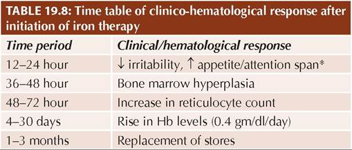
Fig. 19.6: Hemolytic face: (A) Front view; (B) Side view.
Fig. 19.7: Hair end-on appearance on skull X-ray.
and (b) rectangular thickening of metacarpals and phalangeal bones.
Excessive hemolysis may also lead to elevated serum bilirubin and urinary urobilinogen levels.
Antenatal diagnosis is possible by chorionic villous sampling for genetic studies at 10-13 weeks weeks or by fetal blood sampling for HPLC/electrophoresis at 16-20 weeks.
Management of thalassemia aims to (a) maintain optimal Hb levels by repeated blood transfusions to minimize hypoxic damage, prevent marrow expansion and ensure normal growth; (b) prevent iron toxicity due to increased hemolysis and repeated transfusions by chelation therapy; (c) reduce the rate of hemolysis by splenectomy, if necessary, (d) monitor and manage secondary organ dysfunctions including endocrinal complications, and
(e) ultimately, to cure the child with bone marrow transplant and gene therapy. Some important aspects of management are as follows:
a. Transfusion therapy: Repeated blood transfusions to maintain an Hb level at ~10 gm/dl, is the mainstay of treatment in T. major. Super transfusion to maintain Hb levels gt;12 gm/dl is no longer recommended due to higher iron overload.
Most cases require 15-20 ml/kg of packed cell transfusions every 4-5 weeks, which may be given on day care basis. Transfusions should be started as early as possible to prevent expansion of bone marrow and splenomegaly. To avoid transfusion reactions, freshly separated, properly-matched and triple-filtered (to remove granulocytes and plasma proteins) packed cells should be used, along with careful monitoring for transfusion reactions and use of diuretics to avoid overload. Hepatitis B vaccine is advisable in all cases. While transfusion requirements gradually increase with age, sudden increase in transfusion requirements must be investigated for possibility of hypersplenism or aplastic crises.
Newer transfusion modalities include transfusion of specially separated younger cells, i.e. neocytes with longer life span and removal of gerocytes (older cells) by plasmapharesis.
b. Chelation therapy: Iron overload is in direct proportion of the frequency of blood transfusions and degree of hemolysis.
Each 300 ml of packed cells delivers ~200 mg of iron that cannot be excreted by physiological means. Removal of excess iron is as important as transfusion therapy and requires use of iron chelators, starting as early as possible. Three chelating agents are commonly used in India:• Subcutaneous Desferrioxamine (25-50 mg/kg/day) delivered via portable infusion pump over a period of 8-12 hours during night, at least for 5-6 nights/ week. Simultaneous administration of oral vitamin C enhances effectivity of chelation. Important toxic effects are growth failure, cataract, hearing impairment as well as increased risk of yersinia infections.
• Oral Deferiprone (75 mg/kg/day q8hr) is convenient but not as effective as desferrioxamine and usually used as adjuvant to parenteral chelation. Important toxicity of deferiprone is arthropathy, neutropenia and agranulocytosis.
• Oral Deferasirox (20-30 mg/kg/day q24 hr) is the safest chelating agent available at present, though relatively expensive. Common side-effects include gastrointestinal upsets and skin rash. Transient elevation of serum creatinine is common and may require discontinuation of therapy, but progressive renal damage is rare.
Other oral chelators, including a depot preparation of desferrioxamine that needs to be given only twice a week, are under evaluation.
c. Splenectomy to reduce the rate of hemolysis and RBC destruction is indicated only in cases with—(a) transfusion requirements gt;240 ml/kg of packed cells in a year, or (b) massive splenomegaly with signs of hypersplenism or risk of traumatic rupture.
However, it should preferably be deferred till 3-5 years, to cross the infection-prone age. Vaccinations against pneumococci, meningococci, H. influenzae B, etc., and Penicillin prophylaxis are necessary in splenectomized children.
d. Monitoring for secondary organ dysfunctions: Chronic tissue hypoxia, iron overload, extramedullary erythropoiesis and side-effects of transfusions and chelation therapy, invariably leads to significant secondary organ dysfunctions (Table 19.12) in thalassemia major, which must be monitored periodically and managed as required.
TABLE 19.12: Secondary organ dysfunctions in thalassemia
• Endocrinal dysfunction
- Growth retardation (GH deficiency)
- Delayed puberty and hypogonadism
- Hypothyroidism
- T1DM or impaired glucose tolerance
- Hypoparathyroidism
• Cardiac dysfunction
- Congestive cardiac failure
- Arrhythmia
• Hepatic dysfunction
- Progressive cirrhosis
- HBV/HCV infection
• Skeletal involvement
- Osteoporosis
- Pathological fractures
- Arthritis
• Others
- Skin bronzing
- Behavioral issues
e. Bone marrow transplantation (BMT) is best curative option with success rate of ~80-90% if done before significant hemosiderosis develops. Other alternatives for BMT are peripheral stem-cell transplantation and cord blood stem cell transplantation.
f. Other experimental options include:
• Gene therapy with in vitro harvesting of patient's marrow, incorporation of #946;-globin chains (Zynteglo) in auto-stem cells and reinfusion seems to promising option for ultimate cure in future.
• Erythroid maturation agents, e.g. Luspatercept-a recombinant fusion protein that binds TGF-#946; family ligands, blocks the signaling pathway involved in ineffective erythropoiesis and improves erythroid maturation.
Stimulants of HbF synthesis, e.g. hydroxyurea (10-20 mg/kg for 4-30 weeks) or other agents, e.g. butyrates, cytosine A, 5-azacytidine, etc. have marginal role in management of thalassemia to neutralize noxious excess of #946;-chains by increasing #945;-chain production.
Thalassemia has been recognized as a disability under the right of persons with disability act 2016, which entitles them for various benefits, including reservations in educations and jobs.
Prevention of thalassemia involves = (a) pre-marital testing in high-risk populations, (b) genetic counseling in affected persons, and (c) prenatal diagnosis by mutation analysis in amniotic fluid or chorion villus sampling during subsequent pregnancies. This requires establishing the mutation in the previously affected child and carrier parents and use of FISH technique for prenatal diagnosis.
#945;-thalassemia is rare and deletion of all four #945;-globin genes is incompatible with life and presents as hydrops fetalis at birth, since #945;-chains are part of all hemoglobins beyond embryonic period. However, #945;-thalassemia with partial deletion presents either as silent carriers (deletion of one #945;-gene), trait (deletion of two #945;-genes) or HbH disease (deletion of three #945;-genes).
Mixed Hemoglobinopathies: Many other genetic variants of thalassemia often co-exist in some populations with wide clinical spectrum ranging from asymptomatic disease to moderate haemolytic anemia including #948;#946;-thalassemia (partial deletion of both #948;-and #946;-genes), E#946;-thalassemia, i.e. HbE disease (lysine-glutamic acid at 26) with #946;-thalassemia mutation, etc.
19.5.4
More on the topic THALASSEMIA:
- FETAL THERAPY
- 20 Hematologic Disorders of Pregnancy
- Hyposplenic states
- 4 Preconception Counseling and Prenatal Care
- Chapter 7 Genetics and Genetic Disorders in Obstetrics and Gynecology
- Agrawal M.. Textbook of Pediatrics. 3rd ed. — CBS Publishers,2025. — 973 p., 2025
- REFERENCES
- Macrovascular Complications of Diabetes Mellitus
- PRECONCEPTION AND interconception care ^292
- 11 Perinatal Infections