Aging Lesions of the Liver
Rats develop polyploidy, megalokarya, binuclear hepatocytes, intranuclear cytoplasmic invagination, and intracytoplasmic inclusions of hepatocytes that are similar to but not as striking as in the aging mouse.
Although not remarkable in younger animals, the shift to increased ploidy occurs relatively early in life. There is strain-related variation in the incidence of polyploidy in rats. Foci of sinusoidal dilatation and peliosis, either spontaneous or drug-induced, do occur, especially in older animals. Foci of cytoplasmic alteration vary phe- notypically from areas of clearing to acidophilic to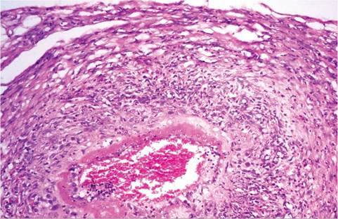
FIG. 2.59. Mesenteric artery from a rat with polyarteritis. There is fibrinoid change in the intima, with inflammation of the media and adventitia.
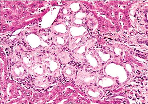
FIG. 2.60. Hepatic portal region from an aged rat, illustrating bile ductular proliferation and fibrosis.
basophilic staining. These changes are of particular interest to the toxicologic pathologist. A striking lesion that is frequently observed in aging rats is bile ductular proliferation. Initially, there are increased numbers of bile ductules in portal tracts, which become progressively dilated, lined by atrophic epithelium, and surrounded by collagenous connective tissue (Fig. 2.60). Extramedullary hematopoiesis may occur in older rats with conditions such as severe chronic renal disease.
Pancreatic Islet Hypertrophy and Fibrosis
Aging rats develop pancreatic islet hypertrophy, which progresses to increasingly severe dissecting fibrosis of pancreatic islets (Fig. 2.61). This lesion has been reported in aging Sprague-Dawley rats, but it occurs in other strains as well.