BOTULISM
ALEKSIJA NEIMANIS
Department of Pathology and Wildlife Diseases, National Veterinary Institute, Uppsala, Sweden
Botulism, also known as limberneck, western duck sickness, alkali poisoning or duck disease, is intoxication by neurotoxin produced by Clostridium botulinum, which acts on the peripheral nervous system to cause progressive flaccid paralysis.
AETIOLOGY
Clostridium botulinum has been classified into seven different types (A-G) based on the antigenically distinct neurotoxin (A, B, C1, D, E, F and G) that each produces. Neurotoxin production occurs only in growing vegetative cells, but it is not released until cells die. Clostridium botulinum types C and D are unique, in that the genes encoding neurotoxin production are located on infectious bacteriophages. Loss of the bacteriophage results in loss of toxigenicity, but toxin-producing potential can be restored with reinfection of the bacterium by the bacteriophage. Only types C and E are significant for wildlife. Type C strains can also produce C2 and C3 toxins, which are not neurotoxic, and their roles in wildlife outbreaks are unknown. An alternate classification scheme based on physiological characteristics divides C. botulinum into four groups. Types C and E are placed in groups III and II, respectively.
Infectious Diseases of Wild Mammals and Birds in Europe, First Edition. Edited by Dolores Gavier-Widen, J. Paul Duff, and Anna Meredith. © 2012 Blackwell Publishing Ltd. Published 2012 by Blackwell Publishing Ltd.
EPIDEMIOLOGY
GEOGRAPHICAL DISTRIBUTION IN EUROPE
Although a well-known cause of wild bird mortality in North America since the beginning of the 20th century, botulism in wild birds was not documented in Europe until the 1960s. The first report of botulism caused by type C1 neurotoxin was in waterfowl in urban ponds in southern Sweden in 1963(1).
Since then, confirmed outbreaks of type C botulism in wild birds have occurred throughout Europe in Austria(2), Czech Republic(3), Denmark(4), France(5), Germany1-6), Hungary1-7), Ireland1-8), Italy(9), the Netherlands(10), Norway(11), Serbia(12), Slovenia(13), Spain(13) and the UK(14). Botulism in birds in Poland is virtually unknown, but a presumptive outbreak of type C botulism in mallards (Anas platy rhynch os) was described in which polymerase chain reaction (PCR) examination detected the type C toxin gene from a bacterial culture of intestine from one duck(15).To date, the only reports of type E botulism in Europe occurred in Friesland province, in the Netherlands, in 1975(10) and in the Canche Estuary of northern France in 1996(16).
Surveys for spores of C. botulinum types C and E in environmental samples demonstrate that spores are widely, but variably, distributed throughout Europe. Although found in soil, they are most prevalent in aquatic sediment. Overall prevalence is generally low, but certain areas are heavily contaminated. Type E spores are especially prevalent in marine sediments (100%) and shores (84%) of the Baltic Sea, the Sound between Sweden and Denmark, the Kattegat and Skagerrak(17). Heavy contamination can also be much more localized. For example, 60% of samples from the Norfolk Broads in Britain contained type E spores(18) compared with 2.7% of samples from aquatic mud collected throughout the UK(19). High concentrations of spores have also been found at sites of previous avian botulism outbreaks (e.g. the Norfolk Broads in the UK(19), British landfill sites(20), the Guadalquivir Basin, Spain(21)). In the Netherlands, 71.6% of mud samples from areas with prior avian botulism had type C spores present(22).
However, high spore prevalence does not necessarily correlate with high incidence of botulism. There are no reports of type E botulism in birds from the heavily spore- contaminated Baltic, whereas only 1 out of 25 (4%) sediment samples from the site of the type E outbreak in Northern France contained type E spores(23).HOST FACTORS
A list of 263 and 31 species known to be affected by type C and type E botulism, respectively, has been published(24). Most bird species, with the possible exception of vultures and other carrion eaters that may have natural and/or acquired resistance, are susceptible to botulism. Age, sex and species composition of affected birds in an outbreak reflect local presence, abundance and feeding habits of birds. Because most cases of botulism occur following ingestion of preformed toxin, feeding habits are the most significant host factor. Depending on the source of toxin in a given outbreak, dabbling ducks that feed at the water’s surface, diving ducks that feed on bottom sediment, shorebirds that feed in shoreline sediment and gulls that feed on carcasses and/or other decaying organic matter have all been particularly vulnerable to type C botulism. Type E botulism occurs in fish- eating birds and can also affect fish. Thousands of birds, primarily black-headed gulls and herring gulls (Larus ridibundus and L. argentatus), died in northern France, presumably after feeding on fish waste(16). Loons, gulls, grebes and occasional waterfowl have suffered from type E botulism in North America. The limited outbreak in the Netherlands occurred in ducks, but the species was not specified. Presence of concurrent fish and bird mortality can indicate type E involvement.
In many incidents of type C botulism in Europe, like in the rest of the world, waterfowl (Anatidae) are the most common species affected, and outbreaks are thought to be perpetuated by the carcass-maggot cycle. However, a different presentation of avian botulism is prevalent in northern Europe.
In the UK(14’25), Ireland1-8) and Sweden1-26), large outbreaks of type C botulism have been reported primarily in gull species (Laridae). In these outbreaks, numerous species of seabirds, particularly herring gulls (Larus argen- tatus), were affected. Epizootics took place along large stretches of coastline, on coastal islands and in estuaries. In the UK and for some gull deaths in Ireland, local landfill and refuse sites were thought to be the source of toxin(8,14), but in no case was the source of toxin definitively determined. Gulls were presumed to have consumed toxin via their scavenging habits. In one incident in the UK, the deaths of 700 gulls were attributed to the spread of contaminated poultry manure onto fields where gulls fed. In Scotland during an outbreak, lesser black-backed gulls (Larus fuscus) were under-represented in the die-off, and one possible explanation was their more maritime feeding habits(25).ENVIRONMENTAL FACTORS
Necessary environmental factors for a botulism outbreak include suitable environmental conditions and appropriate substrate for bacterial growth and toxin production.
Clostridium botulinum type C grows best at temperatures between 30 and 37°C and growth has not been demonstrated at temperatures below 10°C. Most outbreaks therefore occur during the summer and early autumn. In both the UK and the Netherlands, incidence of botulism outbreaks increased during unusually hot summers110’14’27). However, occasional winter and spring outbreaks have been documented in these countries and in the Czech Republic1-28,29’36). Internal temperature in a rotting duck carcass can be significantly higher than ambient temperature, resulting in a suitable microenvironment for toxin production1)30), and type C toxin has been shown to persist through winter in submerged containers1)28) and dead maggots(29).
Both of these factors make outbreaks in colder weather possible. Additionally, some of the Dutch outbreaks occurred near industrial activities that artificially warmed surrounding water(10). By contrast, type E can grow and produce toxin at lower temperatures than type C. Optimal growth occurs between 25 and 30° C, and growth has been documented at temperatures as low as 3.3° C. The outbreaks in France in 1996 took place in February and November and outbreaks in North America often occur during cool autumn weather.Clostridium botulinum requires an anaerobic environment and a high protein substrate for growth. Decaying invertebrate and vertebrate carcasses provide excellent conditions for this. Any environmental factor that results in increased numbers of carcasses and/or allows carcasses to persist can help trigger an outbreak if other necessary factors are present. Examples include sudden changes in water level or water quality, severe weather, chemical spills or pesticide use, limited scavenger access and improper disposal of animal waste.
EPIDEMIOLOGICAL ROLE OF THE AFFECTED SPECIES
Birds dying from botulism provide additional substrate for bacterial growth and can perpetuate an outbreak. Although botulism is an intoxication, expansion of an outbreak via contaminated carcasses can mimic spread of an infectious disease(31). Carcasses also further contaminate the local environment with C. botulinum spores. A large proportion of healthy animals living in an environment contaminated with botulinum spores have spores within their gastrointestinal tracts or other tissues (e.g. 40.5% of animals(10); 50% of mallards(32)). This is of no consequence until the animal dies, regardless of the cause. If conditions are favourable, spores can germinate within the carcass and potentially set off a botulism outbreak. Birds and other animals such as fish can act as mechanical carriers of spores, redistributing spores within the environment.
Gulls in particular are thought to concentrate spores at sites that they are attracted to. This may help to explain higher spore prevalence in landfill and refuse sites versus the general environment in the UK(20). Refuse tested before it was added to landfill sites did not contain detectable spores, further supporting the hypothesis that contamination takes place at the landfill.TRANSMISSION
I n most cases, botulism in wildlife occurs following the ingestion of preformed toxin (food poisoning). Transmission therefore occurs via contaminated food or water. Any decaying organic matter hosting a microenvironment conducive to C. botulinum growth and toxin production may be a potential toxin source. The best- studied scenario is the carcass—maggot cycle of type C botulism in which decomposing carcasses are the microenvironment for C. botulinum proliferation and toxin production, and maggots feeding on these carcasses become contaminated with toxin. Other birds are then exposed to botulinum toxin when they consume these toxic maggots, leading to potentially huge and explosive outbreaks. In one British outbreak, mallards had gorged on maggots shown to contain high levels of botulinum toxin(33). In an outbreak in the Czech Republic necrophagous larvae and pupae of the dipterous flies Lucilia sericata and Calliphora vomitoria contained 80,000 mouse LD50∕g of botulinum toxin(29). Based on experimentally derived toxic doses, a single toxic larva contained enough toxin to kill a duck. They also detected type C toxin in water around carcasses, and from a ptychopterid larva, leeches (Hirudinidae) and sow-bugs (Asellus aquaticus) from within carcasses, but at a much lower concentration (8—800 mouse LD50∕g). In the Salton Sea in California, USA, tilapia fish ( Oreochromis mossam- bicus) were implicated as the source of toxin in a type C outbreak in fish-eating birds, and toxin was detected in the gastrointestinal tracts of sick and dead fish(34). In British and Swedish outbreaks of botulism in herring gulls (Larus argentatus), the scavenging habits of these birds were thought to bring them into contact with the toxin source. In the UK and Ireland, gulls were suspected of ingesting type C toxin at local landfill and refuse sites1-8’20). Type E botulism occurs following ingestion of toxin- containing fish or other marine products.
PATHOGENESIS, PATHOLOGY AND IMMUNITY
Botulinum toxin is the most toxic substance presently known(35) and botulism usually occurs following ingestion of preformed toxin. Oral toxic dose for type C botulism varies greatly between species. In birds, waterfowl are most susceptible, and scavengers and carrion eaters are most resistant. Experimentally, mouse minimum lethal dose per bird varied from 19,000 in teal (Anas crecca) and 64,000 in coots (Fulica atra) to 2,500,000 in crows ( Corvus corone) and black- headed gulls (Larus ridibundus). The mouse LD50 for mallards (Anas platyrhynchos) was 320,000/ bird(36).
In other animals (e.g. foals, poultry), toxico-infectious botulism has been reported. Under the right conditions, spores within the intestinal tract germinate, allowing toxin to be produced i n vivo and absorbed. The other documented form of botulism occurs when a wound is contaminated with spores, and germination and toxin production occur under anaerobic conditions. The significance of these modes of acquisition for wildlife is unknown.
The toxin is absorbed from the gastrointestinal tract and travels via the blood and lymphatic systems to the site of action in the peripheral nervous system. Here, it inhibits acetylcholine release from peripheral cholinergic motor and autonomic nerve endings. Clinical signs in animals are caused by sustained blockage of acetylcholine release at the neuromuscular junction. Toxin does not cross the blood-brain barrier. Uptake of botulinum toxin is closely linked to nerve stimulation, i.e. stimulation facilitates intoxication of the nerve cell(37). The botulinum toxin molecule is composed of a heavy chain and a light chain. A three-step mode of action has been proposed1-35):
1. the neurotoxin binds to the presynaptic cell membrane via the heavy chain
2. the neurotoxin is internalized into the presynaptic nerve cell via endocytosis and the light chain is translocated into the cell cytoplasm
3. the light chain acts as a zinc-endopeptidase to cleave proteins involved in synaptic vesicle docking and fusion to the plasma membrane, thereby blocking the release of acetylcholine at the neuromuscular junction.
This results in the characteristic flaccid paralysis. Each type of botulinum toxin targets a different site on one of three proteins, and type C neurotoxin has two sites of action on two different proteins.
There are no specific gross or microscopic pathologic changes. The acute onset of intoxication means that affected birds typically are in good nutritional condition. Because paralysis limits the ability to eat and drink, birds generally have empty gastrointestinal tracts and are dehydrated. Urates and bright green faeces can distend the cloaca and stain feathers around the vent of affected birds. Waterfowl often have leeches in the eyes, nares and oral cavity.
The binding of botulinum neurotoxin to the presynap- tic membrane is irreversible and the resulting flaccid paralysis is progressive. If intoxication is severe, death results from paralysis of respiratory muscles. If birds are on the water when intoxicated, paralysis of neck muscles can cause death by drowning. In other cases, paralysis leads to death by exposure to the elements, dehydration, starvation or predation.
Duration of botulinum toxin action is not known in animals. However, in humans, time to complete recovery is typically 2-4 months at the neuromuscular junction, but can take more than a year in the autonomic nervous system(37).
Botulinum neurotoxin is so toxic that, for most species, the dose required to develop acquired immunity through natural exposure is higher than the lethal dose(38). No neutralizing antibodies were found in mallards given multiple sublethal doses of botulinum toxin type C(36). Naturally occurring antibodies in captive ducks suffering from botulism 2 weeks after onset of disease were detected, but
these disappeared by 5 weeks1-39). Immunitywas short-lived and not protective against future exposure. However, in a study of free-living scavengers and carrion-eaters, 90% of turkey vultures (Cathartes aura), 42% of crows (Corvus brachyrhynchos), 25% of coyotes (Canis latrans) and 17% of Norway rats (Rattus norvegicus) sampled had naturally occurring antibodies to various types of C. botulinum toxin(38). Similarly, although ring-billed gulls (Larus dela- warensis) are susceptible to botulism type E, antibody production was experimentally induced in gulls fed lower levels of toxin(40). Sublethal natural exposure in the wild may induce some degree of immunity. Vaccination of wild birds against botulism type C with commercial mink vaccine(41) improved survival in challenge studies, but in most cases vaccination of free-ranging birds is not warranted.
CLINICAL SIGNS AND TREATMENT
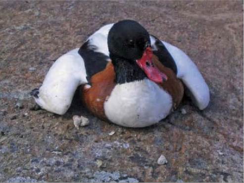
Figure 36.1 A common shelduck (Tadorna tadorna) moderately affected by botulism type C. Wings are dropped at the sides and the duck has lost the use of its legs, but it remains alert and responsive.
The hallmark sign of botulism is progressive, flaccid paralysis of skeletal muscles. The rate of onset and the severity of the clinical signs reflect the amount of toxin ingested. In early stages, birds have difficulty flying or can only fly short distances. Once flight capability is lost, birds can still often stand and walk, although gait may be impaired or uncoordinated. Eventually, they become recumbent, but may be able to propel themselves forward with their wings. While resting, wings can be dropped away from the body (Figure 36.1). In late stages, birds are prostrate and neck, head and respiratory muscles are affected. Inability to support the head results in ‘limberneck’, described in ducks and other species (Figure 36.2). Breathing becomes laboured, and the nictitating membrane may be prolapsed. Until birds reach the final stages, they generally are bright and alert, despite the paresis and paralysis.
Treatment of affected birds can be highly effective (75— 90% recovery rate(24)). Mildly to moderately affected birds can be treated simply by providing food, water and shelter from the elements and predators. Antitoxin has been given in some cases, but toxin already bound to and taken up by nerve cells cannot be neutralized. Recovered birds have no protective immunity so must not be released back into the site of an ongoing outbreak. Vaccination of birds prior to release can help reduce re-intoxication. Treatment requires a large number of resources and may only be warranted in certain situations (e.g. if threatened or endangered species are involved).
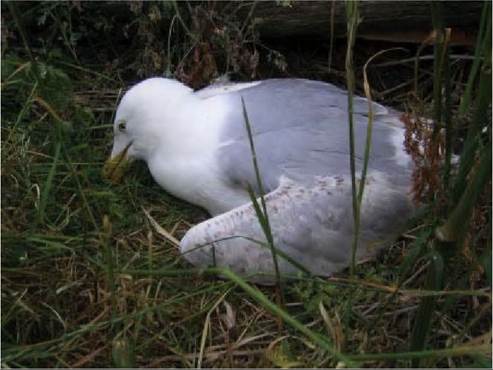
Figure 36.2 A herring gull (Larus argentatus] severely affected by botulism type C. The bird is prostrate and the neck muscles are paralyzed (limberneck).
DIAGNOSIS
Definitive diagnosis of botulism is challenging and disease often diagnosed presumptively based on clinical signs coupled with the absence of gross and microscopic lesions and negative test results for other aetiologies. Toxin proliferation within a decaying carcass can occur post mortem, resulting in false positives, so the ideal sample for confirmatory testing is serum from sick birds. Because the majority of toxin can be fixed at the site of action in the peripheral nervous system and result in false negatives, it is best to test a number of birds in a given outbreak. Serum can be frozen until tested, but toxin activity significantly decreases over time.
Botulinum toxin is a protein, so the ideal confirmatory test must detect the presence of biologically active proteins. Currently, the mouse bioassay is the only test available for type C botulism that meets these requirements. Functional in vitro assays for type E are under development. Protocols for the mouse test vary, but briefly, serum samples (0.5— 1 ml) are injected into two groups of mice. One group receives protective antitoxin 30 minutes before serum injection. The test is considered positive if unprotected mice show clinical signs of botulism or die and protected mice remain unaffected throughout the test. Onset of clinical signs can be delayed if toxin concentrations within the sample are low, so mice are observed for 4 days. Post mortem examinations can be carried out on any dead mice to ensure that no other aetiologies (e.g. bacterial infection with Pas- teurella multocida) were responsible.
For a variety of reasons, including the terminal use of animals, cost, and time needed to reach a diagnosis, numerous other diagnostic tests have been developed. Tests for human botulism have been reviewed and com- pared(35). Other diagnostic tests used for wildlife include immunoassays (e.g. enzyme-linked immunosorbent assay (ELISA)) and molecular techniques (PCR). ELISA tests, although more rapid, are not as sensitive as the mouse test. An improved test, an antigen- capture ELISA for type C botulism that concentrates toxin present within a sample, has been developed(42). At sample volumes less than 1 ml of serum, the mouse bioassay is still more sensitive. However, at these volumes, this ELISA is a good screening test if three or more animals from a given outbreak are tested. If sample volumes greater than 1 ml are used, this ELISA may be as sensitive or more sensitive than the mouse test. Unfortunately, this volume of serum often is difficult to obtain from birds in natural outbreaks.
PCR techniques have been developed to detect the botulinum neurotoxin gene, but these tests are best employed to detect the presence of spores in the environment. Caution should be used when interpreting toxin production potential from environmental samples using PCR, because this technique does not detect activity of the gene. Additionally, botulinum spores often are present in healthy animals, so detection of the toxin gene in samples from animals by PCR does not mean that the animal died from botulism. A PCR technique to detect the type C toxin gene in only active cells provides stronger evidence that the animal died from botulism(43).
A promising i n vitro assay is the endopeptidase- mass spectrometry assay under development for botulism diagnosis in humans(44). This functional assay takes advantage of the specific enzymatic activity of each botulinum toxin. Cleaved protein products are then detected with highly sensitive mass spectrometry, and toxin type is determined based on product size. This assay is designed to detect human botulism, i.e. only types A, B, E and F, and requires further refinement.
MANAGEMENT, CONTROL AND REGULATIONS
The management and control of botulism are difficult, but the most effective way to mitigate losses from botulism in wildlife is through prevention of outbreaks. Because decaying organic matter can serve as a substrate for C. botulinum growth and toxin production, environmental management should focus on minimizing die-offs of invertebrates and vertebrates that overwhelm local scavenger capacity. Suggested strategies include avoidance of sudden changes in water levels of managed wetlands, removal of physical hazards for birds, avoidance of practices resulting in fish kills, delay of hunting until cooler weather and promotion of scavenging activity(31). Excluding susceptible birds from an affected area during an outbreak has been attempted. Removal of carcasses during an epizootic should, in theory, be an effective way to eliminate substrate for further bacterial growth and toxin production. However, the effectiveness of botulism control via carcass pick- up during an outbreak is questionable. In one study, on average only 32.1% of marked carcasses were detected, and larger species were over-represented in carcass collection(45). Small, open, easily accessible environments may be more suitable to this type of control. Finally, landfill site management to limit attractiveness for birds may help to prevent botulism in gulls.
Botulism in humans, but not wildlife, is notifiable at the EU level (Decision no. 2119/98/EC of the EU Parliament and Council).
PUBLIC HEALTH CONCERN
Humans are susceptible to type E botulism, so care should be taken when handling tissues and samples from birds that died from type E. Type C neurotoxin does not affect humans.
SIGNIFICANCE AND IMPLICATIONS FOR ANIMAL HEALTH
Botulism in wildlife has little significance for domestic species. One outbreak of botulism type C in cattle was linked to a waterfowl die-off in a wetland in Canada where cattle were grazing and drinking(46), but this is unusual. Conversely, botulism is one of the most significant diseases of migratory birds, especially waterfowl and shorebirds, worldwide. Huge die-offs of hundreds of thousands or even a million birds have been reported in North American waterbirds. In Europe, die-offs have not been as dramatic, but some outbreaks have claimed thousands of waterfowl and gulls. The largest reports include 60,600 and 70,000 birds in the Netherlands in 1976 and 1983, respectively1-10), at least 10,000 gulls in France in 1996(16), 4,040 birds, primarily gulls, in the UK in 1975(14) and probably thousands of seabirds, primarily herring gulls, in Sweden from 2000 to 2004(26). Additionally, botulism outbreaks may negatively impact threatened and endangered species. For example, avocets (Recurvirostra avosetta), a critically endangered species in the Czech Republic, were involved in a botulism outbreak there(47). Other rare or endangered birds killed by botulism in the Czech Republic included spoonbills (Platalea Ieucordia), curlews (Numenius arquata), black-tailed godwits (Limosa limosa) and redshanks ( Tringa totanus). Finally, although botulism can be the proximate cause of death of wildlife, the ultimate cause of the epizootic (e.g. the initiating conditions that caused the increased numbers of carcasses initially or the environmental change that facilitated C. botulinum germination and toxin production) may be of the most significance. Thus, changes in patterns and prevalence of botulism outbreaks may signal underlying environmental perturbation.
More on the topic BOTULISM:
- Deep Frostbite
- 5 Appendices
- THE CLOSTRIDIAL DISEASES
- Nerve Conduction Studies
- Clostridium difficile and Clostridium perfringens: Clostridial Enteropathy
- TECHNICAL FACTORS OF NEEDLE ELECTROMYOGRAPHY
- Pregnant Women and Chemical-Biological Warfare