Epidemiology of BTB Caused by M. bovis in Burkina Faso
Evidence of the presence of BTB in Burkina Faso dates back to the colonial period. There are often differences in the prevalence detected by tuberculin skin test surveillance and data obtained from abattoirs because the source of animals differs for the two datasets.
In Bobo-Dioulasso, based on the single cervical intradermal test (SIT), the prevalence was estimated to be 10% in 1951 (n = 390) and 12.6% in 1952 (n = 111) (Sere 1966). A large survey conducted in 1969 in Dori and Dedougou using SIT reflected a prevalence of 6.0 and 2.8%, respectively (Gidel et al. 1969b). Follow-up studies conducted in Bobo-Dioulasso in 1999 confirmed the persistence of BTB with an estimated prevalence of 13.0% (Vekemans et al. 1999). There seems to be significant differences in the prevalence of BTB depending on the farming system. According to a study conducted in an intra-urban dairy farm in Ouagadougou in 2004, the BTB prevalence was estimated to be 27.7% (Traore et al. 2004). In intra-urban and suburban farms in Ouagadougou representing three different beef production systems (extensive, an enhanced system, including semi-intensive and intensive farming, and an intra-urban-intensive breeding variant), the BTB prevalence in 2012 was 5.1, 3.0, and 14.8%, respectively, for animals originating from the three systems (Boussini et al. 2012).
Because of the lack of funding to conduct intradermal tuberculin tests, BTB is usually diagnosed in Burkina Faso by detecting suspected tuberculous lesions during meat inspection in abattoirs. Trained animal health professionals (meat inspectors) conduct these inspections, and the data that they gather have been the subject of several published reports (Sere 1966; Gidel et al. 1969a). Based on routine meat inspections conducted at Bobo-Dioulasso slaughterhouse in 1995 and 1999, the BTB prevalence was, respectively, 3.7 and 19% (Delafosse et al.
1995; Vekemans et al. 1999).Several factors appear to influence the prevalence and distribution of BTB in Burkina Faso. These include husbandry systems, the age and sex of the cattle, and uncontrolled trans-boundary movement of livestock from neighboring BTB-infected countries (Gidel et al. 1969b; Tarnagda et al. 2014; Regnoult 1963). The prevalence of BTB is higher in farming systems where animals live in close contact with one another, where large numbers of cattle are housed in a confined airspace and where there is inadequate health monitoring (Vekemans et al. 1999; Boussini et al. 2012), thus partly explaining why the Sahelian regions generally have a higher prevalence of BTB (Gidel et al. 1969b). The association between a longer lifespan of an animal and the increased risk of having BTB (Vekemans et al. 1999) and the animal’s sex is reflected by the prevalence in females of 6.7% compared to 2.9% in males and 17% in 2-year-old animals compared to 33.7% in those older than 6 years (Boussini et al. 2012; Tarnagda et el. 2014).
Burkina Faso is located in the heart of West Africa, and it is a transit hub for the movement of people and their livestock to and from its six neighboring countries
Table 11.2 Mycobacterium bovis spoligotype signatures isolated from humans and animals in Burkina Faso (Sanou et al. 2014)
| SB numbersa | Host species | Geographical location | Clonal complex (%)b |
| SB2282 | Cattle | Bobo-Dioulasso | Af5 (3%) |
| SB2283 | Human | Solenzo | Af5 (3%) |
| SB1398 | Cattle | Bobo-Dioulasso | Af5 (6%) |
| SB2284 | Human | Bobo-Dioulasso | Af5 (3%) |
| SB2285 | Cattle | Bobo-Dioulasso | Af5 (3%) |
| SB0857 | Cattle | Ouagadougou | Af1 (3%) |
| SB0300 | Cattle, human | Bobo-Dioulasso, Ouagadougou | Af1 (9%) |
| SB2286 | Cattle | Ouagadougou | Af1 (9%) |
| SB2287 | Cattle | Ouagadougou | Af1 (3%) |
| SB0944 | Cattle, human | Bobo-Dioulasso, Ouagadougou, Koupela | Af1 (52%) |
| SB1439 | Cattle | Ouagadougou | Af1 (3%) |
| SB2288 | Human | Ouagadougou | Af1 (3%) |
| Total | Af5 (18.2%) | ||
| Af1 (81.8%) | |||
aSB number = name of spoligotype based on M.bovis.org database nomenclature bAf1 = African 1 clonal complex; Af5 = putative African 5 clonal complex
(Sere 1966; Sanou et al. 2014).
Thus, cross-border cattle movement, particularly from Mali and Niger, appears to significantly influence the epidemiology of BTB, and it is likely to have caused an increase in the prevalence of BTB in Burkina Faso. This assumption is supported by data from local abattoirs in the Nouna sector that is adjacent to the BTB-infected areas of Mopti, Segou, and Macina in Mali (Sanou et al. 2014; Regnoult 1963).Mycobacterium bovis is the major causative agent of BTB and is responsible for 89-97% of cases in cattle in Burkina Faso (Letroteur 1952; Sere 1966). There are many gaps in the understanding of the epidemiology of BTB in Burkina Faso, but a detailed molecular epidemiological investigation of the types of human and bovine M. bovis isolates in 2014 shed some light on the origin of the infection in the country. The strains of M. bovis in Burkina Faso are characterized by a considerable genetic diversity, and at least 12 M. bovis spoligotypes, divided into two subgroups, were identified (Table 11.2).
All M. bovis strains in this study belonged either to the Af1 clonal complex, found in other countries of West-Central Africa, such as Mali, Cameroon, Nigeria, Chad, and Niger, or to the putative Af5 clonal complex that has also been described in Mali. In Burkina Faso, the putative Af5 strains comprise about 18.2% of the isolates and are geographically localized in the western region (Bobo-Dioulasso and Solenzo), an area bordering Mali (Fig. 11.1). The presence of common or related genotypes in Burkina Faso and Mali is probably the consequence of transhumance between these countries, and of the passage, through Burkina Faso, of Malian
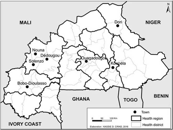
Fig. 11.1 Map of Burkina Faso showing major cities and regional and international borders
livestock to Ghana and Nigeria in the south. Spoligotype signatures belonging to the putative Af5 clonal complex have been reported in Europe, and it is likely that the putative Af5 clonal complex was introduced into this region of Africa from Europe, probably via North Africa (Muller et al.
2008; Haddad et al. 2001; Sahraoui et al. 2009). The larger group of isolates, comprising about 81.8% of the total national M. bovis isolates, belongs to the Af1 clonal complex, also including the SB0944 spoligotype pattern, a dominant strain that accounts for almost half of all the isolates. This group is characterized by the lack of spacer 30 and is also the most common type in other West-Central African Sahelian countries such as Mali, Nigeria, Chad, Niger, and Cameroon (Boukary et al. 2012; Muller et al. 2009). In the Af1 clonal complex, the SB0944 spoligotype signature is considered the most recent common ancestor (progenitor). It is the most common pattern in this group and comprises 40% of isolates in Chad, 46.1% in Nigeria, and 62.7% in Cameroon. It is also the most abundant type (52%) in Burkina Faso (Sanou et al. 2014; Muller et al. 2008). The spread of the Af1 clonal complex over this large area of West-Central Africa is probably caused by the long-distance, cross-border migration of livestock, a feature of the transhumant production system mainly practiced by the Fulani pastoralists in the Sahel (Muller et al. 2008).11.3
More on the topic Epidemiology of BTB Caused by M. bovis in Burkina Faso:
- Control of BTB in Burkina Faso
- Other Mycobacteria as the Cause of BTB in Burkina Faso
- Chapter 11 Bovine Tuberculosis: Status, Epidemiology, and Public Health Implications in Burkina Faso
- Tuberculosis Caused by M. bovis in Humans in Uganda
- The Use of Molecular Epidemiology in Understanding the Dynamics of M. bovis
- The Epidemiology of BTB in Malawi
- Molecular Epidemiology of M. bovis in Humans and Other Hosts in Africa
- The Epidemiology of BTB in Cattle in Cameroon
- Prevalence and Epidemiology of BTB in Rwanda
- Molecular Epidemiology of BTB in Nigeria
- Chapter 22 Holes and Patches: An Account of Tuberculosis Caused by Mycobacterium bovis in Uganda