GENETIC DISORDERS
Considering the large number of relatively inbred breeds of rabbits, it is not surprising that rabbits may be afflicted with a number of hereditary disorders. A few disorders that are likely to be encountered, particularly in laboratory rabbits, are summarized in the following sections.
Congenital Glaucoma: Buphthalmia
This condition occurs most frequently in New Zealand White rabbits. Buphthalmia is characterized clinically by enlargement of 1 or both eyes, with subsequent corneal opacity. Abnormalities may occur within the first few weeks of life, but usually they are first evident by 3-5 months of age. The primary defect has been identified as an absence or underdevelopment of the outflow channels, with incomplete cleavage of the iridocorneal angles. With impaired drainage of aqueous humor from the anterior chamber, the increased intraocular pressure results in megaloglobus, increased corneal diameter, and protrusion of the corneal contours (Fig. 6.97). The sclera is relatively immature at this stage and thus expands to accommodate the increased volume of aqueous humor within the globe. The defect is inherited as an autosomal recessive allele, with incomplete penetrance. Therefore, some animals that are homozygous for the bu/bu gene may show no evidence of the disease. Buphthalmia does not appear to cause discernable discomfort in affected animals.
Anterior Corneal Dystrophy
Although rarely noticed or reported, anterior corneal dystrophy has been described in a closed colony of Dutch Belted rabbits, but is also seen by others in this
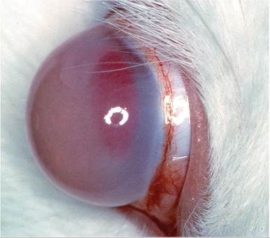
FIG. 6.97. Buphthalmia in a New Zealand White rabbit. There is marked distention of the cornea of the affected eye.
breed. Affected rabbits had unilateral or bilateral focal linear or plaque-like epithelial and subepithelial opacities of the central and paracentral corneas.
Basement membranes were thickened and irregular, and adjacent stroma revealed disorganized collagen lamellae. Epithelium was often thin and disorganized in affected areas. Corneal dystrophy has also been reported in New Zealand White rabbits, but may differ in that epithelium and basement membranes were normal. These syndromes, particularly the former, are likely to have a genetic basis. They are of import for this text because both breeds are commonly used as laboratory animals, and such lesions have consequences for toxicology research. Lipid keratopathy is also common in hyperlipidemic rabbits, which has a genetic basis in the Watanabe rabbit with heritable hypercholesterolemia (see Atherosclerosis/Hypercholesterolemia and "Xanthoma- tosis/Hypercholesterolemia”).Ocular Cataracts
Spontaneous cataracts are a common finding in rabbits of various breeds. Fetal and congenital cataracts have been reported, and cataracts are known to occur in association with Encephalitozoon cuniculi infection of the lens and anterior segment of the eye. A recent study examined the eyes of NZW and NZW ? New Zealand Red F1 hybrid rabbits and found that the incidence of cataracts was consistent with an autosomal recessive genetic disorder in NZW rabbits, with a significantly lower incidence in hybrids. Males and females were equally affected, and there was no indication that incidence increased with age. The primary differential diagnosis is cataracts associated with E. cuniculi.
Malocclusion
Maxillary brachygnathia (erroneously termed mandibular prognathism) is the most common genetic disorder in domestic rabbits. The defect is inherited as an autosomal recessive trait. Normally, occlusion brings the lower
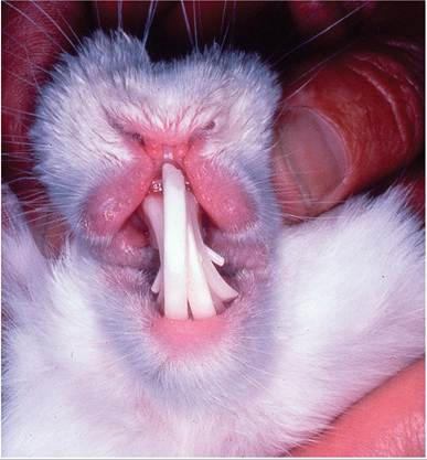
FIG. 6.98. Overgrowth of incisor and peg teeth in a New Zealand White rabbit due to genetically inherited brachygnathism of the maxillary bones.
incisor teeth into apposition against the upper secondary incisors (peg teeth) located behind the large upper incisors.
In rabbits with maxillary brachygnathia, the maxilla is abnormally short relative to the mandible. The overshot lower j aw results in misalignment, failure of the incisors to wear normally, and impaired mastication. In domestic rabbits, growth of the combined lengths both the upper and lower incisors has been shown to be over 20 cm/year. Thus, malocclusion will cause overgrowth of the incisors in a relatively short period of time (Fig. 6.98). Although not as evident as malocclusion of the incisors, malocclusion of premolar and molar teeth with overgrowth also occurs in rabbits. Dietary deficiencies may also be a contributing factor. Overgrowth with ridging of the incisors and distortion of the cheek teeth has been observed in rabbits fed a diet deficient in calcium and vitamin D.Splay Leg
Preweanling rabbits may develop a condition in which the front legs, hind legs, or all 4 legs splay to the side (Fig. 6.99). It is generally believed that the condition has a recessive genetic basis, but it involves various breeds of rabbits. It is exacerbated when kits are raised on substrates that do not provide adequate traction, and can be ameliorated if traction is provided. Morphologic changes in the hind legs include coxofemoral subluxation, shallow acetabula, lateral patellar luxation, valgus deformity, and bowing of the tibia.
Megacolon-Syndrome of Spotted Rabbits
The megacolon-syndrome is associated with an incomplete dominant mutant allele of the English spotting locus (En) that determines spotted coat phenotype in breeds such as the English Spot and the Checkered Giant when the allele is homozygous. The syndrome can affect
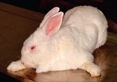
FIG. 6.99. Splay leg in a preweanling rabbit. Affected rabbits are otherwise normal and the condition can be improved by housing the rabbit on more tractable surfaces. (Source: S. Vandewoulde, Colorado State University, Fort Collins, CO.
Reproduced with permission from S. Vandewoude. )any rabbit with the spotted phenotype carrying this mutation. Albino rabbits with this gene mutation do not manifest the coat color phenotype, but can suffer from the disease. Homozygous rabbits are "subvital" due to a megacolon-syndrome, which is not congenital, but rather progresses with age of the rabbit. The pathophysiology has not been extensively investigated, but affected rabbits appear to have a sodium absorption defect in the cecum and 1 study noted a relative hypogangliosis in the distal parts of the gut. Rabbit fanciers refer to this as "cow pie syndrome" since fecal pellets are much larger and poorly formed and cecotrophs are atypically large and torpedo-shaped.
Polycystic Kidney Disease
Polycystic kidney disease was reported in adult New Zealand White rabbits from multiple commercial sources. Cysts were found within the cortex, less than 2 mm in diameter, and often not noted grossly. Microscopically, dilation of Bowman's space and/or proximal tubules, irregular thickening and splitting of basement membranes, and expansion of cortical and medullary interstitium were observed. This syndrome was suspected to have a genetic basis. An autosomal recessive inheritance of cortical renal cysts was noted in partially inbred IIIvo rabbits, which had featured single to several hundred small cortical cysts, which arose in rabbits after 1 month of age.
BIBLIOGRAPHY FOR GENETIC DISORDERS
Congenital Glaucoma: Buphthalmia
Burrows, A.M., Smith, T.D., Atkinson, C.S., Mooney, M.P., Hiles, D.A., & Losken, H.W. (1995) Development of ocular hypertension in congenitally buphthalmic rabbits. Laboratory Animal Science 45:443-444.
Hanna, B.L., Sawin, P.B., & Sheppard, L.B. (1962) Recessive buph- thalmos in the rabbit. Genetics 47:519-529.
Tesluk, G.C., Peiffer, R.L., & Brown, D. (1982) A clinical and pathological study of inherited glaucoma in New Zealand White rabbits.
Laboratory Animals 16:234-239.Anterior Corneal Dystrophy
Moore, C.P., Dubielzig, R., & Glaza, S.M. (1987) Anterior corneal dystrophy of American Dutch Belted rabbits: biomicroscopic and histopathologic findings. Veterinary Pathology 24:28-33.
Port, C.D. & Dodd, D.C. (1983) Two cases of corneal epithelial dystrophy in rabbits. Laboratory Animal Science 33:587-588.
Ocular Cataracts
Gelatt, K.N. (1975) Congenital cataracts in a litter of rabbits. Journal of the American Veterinary Medical Association 167:598-599.
Munger, R.J., Langevin, N., & Podval, J. (2002) Spontaneous cataracts in laboratory rabbits. Veterinary Ophthalmology 5:177-181.
Weisse, I., Niggeschultz, A., & Stotzer, H. (1974) Spontane, congenitale Katarakte bei Ratte, Maus und Kaninchen. Archives of Toxicology 32:199-207.
Malocclusion
Fox, R.R. & Crary, D.D. (1971) Mandibular prognathism in the rabbit: genetic studies. Journal of Heredity 62:23-27.
Verstraete, F.J.M. (2003) Advances in diagnosis and treatment of small exotic mammal dental disease. Seminars in Avian and Exotic Pet Medicine 12:37-48.
Zeman, W.V. & Fielder, F.G. (1969) Dental malocclusion and overgrowth in rabbits. Journal of the American Veterinary Medical Association 155:1115-1119.
Splay Leg
Arendar, G.M. & Milch, R.A. (1966) Splay-leg—a recessively inherited form of femoral neck anteversion, femoral shaft torsion and subluxation of the hip in the laboratory lop rabbit: its possible relationship to factors involved in so-called “congenital dislocation” of the hip. Clinical Orthropaedics and Related Research 44:221-229.
Innes, J.R.M. & O'Steen, W.K. (1957) Splayleg in rabbits: an inherited disease analogous to joint dysplasia in children and dogs. Laboratory Investigation 6:171-186.
Joosten, H.F.P., Wirtz, P., Verbeek, H.O.F., & Hoekstra, A. (1981) Splayleg: a spontaneous limb defect in rabbits—genetics, gross anatomy, and microscopy. Teratology 24:87-104.
Owiny, J.R., Vandewoude, S., Painter, J.T., Norrdin, R.W., & Veer- machaneni, D.N.R. (2001) Hip dysplasia in rabbits: association with nest box flooring. Comparative Medicine 51:85-88.
Megacolon-Syndrome of Spotted Rabbits
Bodeker, D., Turck, O., Loven, E., Wieberneit, D., & Wegner, W. (1995) Pathophysiological and functional aspects of the megacolon-syndrome of homozygous spotted rabbits. Zentralblatt Veterinarmedizin A 42:549-559.
Gerlitz, S., Wessel, G., Wieberneit, D., & Wegner, W. (1993) The problems of breeding spotted rabbits. 3. Variability of the pigmentation grade, ganglionic intestinal wall supply, relationship to pathogenesis-animal breeding and animal welfare aspects. Deutsche tierarztliche Wochenscrift 100:237-239.
Wiebernett, D. & Wegner, W. (1995) Albino rabbits can suffer from megacolon-syndrome when they are homozygous for the “English Spot” gene (En/En). World Rabbit Science 3:19-26.
Polycystic Kidney Disease
Fox, R.R., Krinsky, W.L., & Crary, D.D. (1971) Hereditary cortical renal cysts in the rabbit. Journal of Heredity 62:105-109.
Mauer, K.J., Marini, R.P., Fox, J.G., & Rogers, A.B. (2004) Polycystic kidney syndrome in New Zealand White rabbits resembling human polycystic kidney disease. Kidney International 65:482-489.