NEOPLASMS
Rabbits develop numerous types of neoplasia, details of which can be found in the General References. Common tumors that are likely to be encountered are summarized in the following sections.
Uterine Adenocarcinoma
Uterine adenocarcinoma is the most commonly encountered spontaneous neoplasm occurring in O. cuniculus. The relatively low incidence of this tumor seen in most commercial rabbitries and research facilities is due to the fact that these animals are usually relatively young. In 1 study, the incidence of uterine adenocarcinoma in does 2-3 years of age was around 4%, and in does 5-6 years of age, around 80%. Thus, there is a striking increase in the incidence of uterine tumors with increasing age. A variety of breeds are affected. Cystic endometrial hyperplasia is often found in the rabbit uterus, and believed to be antecedent to neoplastic progression.
On gross examination, the tumors appear as nodular, frequently multicentric masses that often involve both uterine horns (Fig. 6.100). On the cut surface, masses are firm, frequently with a cauliflower-like surface and central necrosis. Serosal implantation and metastases to the lung (Fig. 6.101) and liver often occur. Typical microscopic changes of the tumor are those of an adenocarcinoma, with invasion of the underlying layers forming acinar and tubular structures (Fig. 6.102). In rapidly growing tumors, necrotic areas are frequently observed. Metastases and tumor implants are similar to the primary neoplasm, often with a prominent stromal component.
Lymphosarcoma: Lymphoma
Neoplasms of lymphocytic origin are the most common malignancy encountered in juvenile and young adult rabbits. Anemia, low hematocrit, and terminally elevated BUN (due to kidney involvement) are typical changes seen clinically. Leukemia occasionally occurs, particularly during the terminal stages of the disease.
In 1 rabbit strain, an autosomal recessive gene has been implicated as a factor in susceptibility to the disease. Others have speculated on the relationship of rabbit endogenous retrovirus (cause and effect never proven).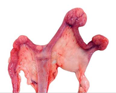
FIG. 6.100. Uterine adenocarcinomas in an aged doe. These tumors are often multicentric, involving both uterine horns. (Source: D. Imai, University of California, Davis, CA. Reproduced with permission from D. Imai.)
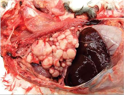
FIG. 6.101. Metastatic uterine adenocarcinoma in the lung of an aged doe. Uterine adenocarcinomas frequently metastasize to the lungs. (Source: B.G. Caserto, Cornell University, Itaca, NY. Reproduced with permission from B.G. Caserto.)
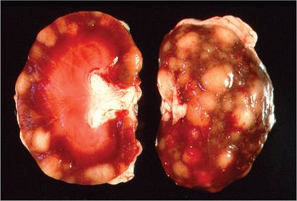
FIG. 6.103. Kidneys from a young rabbit with lymphosarcoma. Multiple pale nodular masses are visible in the cortex, a frequent finding in lapine lymphosarcoma.
Lymphosarcoma in the rabbit has unique patterns of organ involvement. Gross findings usually include pale kidneys with irregular cortical surfaces, enlarged GALT and mesenteric lymph nodes, hepatosplenomegaly, and patches of pale bone marrow. Nodular masses in the subcutis, lung, and eye involvement may occur. The most pathognomonic feature is renal involvement. On cut surface, changes are typically confined to the renal cortices (Fig. 6.103). Involvement of the GALT (pharyngeal tonsils, stomach mucosa, Peyer's patches, appendix, mesenteric lymph node, etc.) is frequent (Fig. 6.104). The wall of the stomach may be markedly thickened, with irregular surface plaques and mucosal ulceration. Examination of bone marrow will often reveal involvement (Fig. 6.105). The liver and spleen are often enlarged, pale, and swollen. Enlarged peripheral lymph nodes and pulmonary nodules may also be encountered.
On histopathology, there are diffuse infiltrates of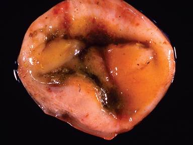
FIG. 6.104. Diffuse mural thickening of the cecal appendix in a rabbit with lymphosarcoma. Lymphosarcoma most frequently arises in gut-associated lymphoid tissue in rabbits. (Source: D. Imai, University of California, Davis, CA. Reproduced with permission from D. Imai.)
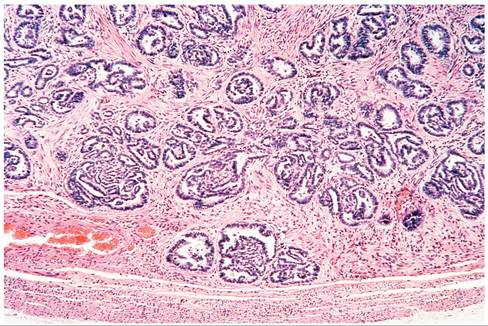
FIG. 6.102. Uterine adenocarcinoma from an aged New Zealand White doe. Note the invasive glandular structures lined by neoplastic epithelium.
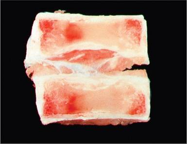
FIG. 6.105. Vertebral bone from a rabbit with lymphosarcoma. Note the pale bone marrow due to neoplastic involvement. The bone marrow is a frequent site of involvement in lapine lymphosarcoma, but is seldom examined.
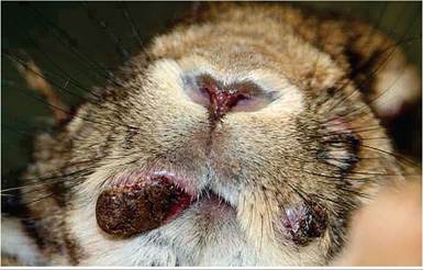
FIG. 6.106. Cutaneous nodules involving the muzzle of a rabbit with cutaneous lymphoma. These multicentric tumors are populated by pleomorphic cells infiltrating the dermis without epitheliotropism, and arise on various sites of the body. (Source: Ritter et al. 2012. reproduced with permission from SAGE Publications.)
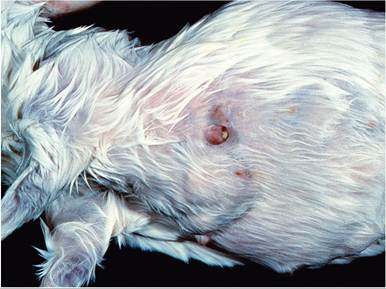
FIG. 6.107. Mammary adenocarcinoma in an aged New Zealand White doe.
lymphoblastic cells in the affected regions of stomach or intestine and in the interstitial regions of the renal cortex, with distortion of the normal architecture and relative sparing of the glomeruli. In the liver, there are periportal to diffuse sinusoidal infiltrates of neoplastic cells. Diffuse infiltration may occur in the spleen, lymph nodes, bone marrow, uveal tract, adrenal gland, and ovary. B-cell, T-cell, and mixed lymphoid cell populations have been documented.
Diffuse large B-cell and T-cell-rich B-cell lymphomas, presenting as one or more nodules (Fig. 6.106) on various locations of the body, have recently been reported in adult pet rabbits of various breeds in Europe, but not domestic rabbits from North America. The subcutis and dermis were infiltrated, but epitheliotropism was not observed. Involvement of other organs was variable. Neoplastic infiltrates were described as highly pleomorphic and often contained multinucleated giant cells. These differ from epitheliotropic T-cell lymphomas, which have been associated with exfoliative dermatosis in rabbits. In such cases, neoplastic cells infiltrated both the dermis and epidermis, with infiltration of distant organs.
Thymoma
Thymomas are uncommon, but well represented among neoplasms in domestic rabbits. They arise in rabbits between 1 and 4 years of age, and are often found as incidental findings at necropsy. Rabbits may present with dyspnea due to an anterior mediastinal mass. Rarely, thymomas may become metastatic. More commonly, paraneoplastic syndromes may arise. Concurrent hypercalcemia, exfoliative dermatosis, and periodic exophthalmos have all been reported as paraneoplastic syndromes associated with thymomas in rabbits.
Mammary Adenomas and Carcinomas
Glandular tumors of the mammary gland (Fig 6.107) are relatively frequent in multiple breeds of rabbits, including laboratory rabbits, arising around 3-4 years of age.
The majority of mammary tumors are carcinomas, including tubular, papillary, tubulopapillary, solid, adenosquamous, comedo, complex, ductal, cribriform, anaplastic, and spindle cell carcinomas. Cystic mastitis is conjectured as a prelude to neoplastic transformation from benign adenomas to adenocarcinomas. Metastasis to regional lymph nodes and lungs, as well as other organs, has been reported.
Bile Duct Adenomas and Adenocarcinomas
Bile duct tumors are the fourth most common reported tumor in rabbits. They are usually found incidentally at necropsy in older rabbits and appear as solitary or multiple cystic lesions filled with thick yellow to tan fluid.
Pseudo-odontoma: Odontoma
Odontomas are tumors of odontogenic origin that feature both epithelial and mesenchymal cells of tooth origin that are well differentiated (Fig. 6.108). When the cells within these masses are disorganized to the extent that they do not resemble a tooth, they are termed complex odontomas. Such tumors arise rarely from the elodont tooth roots of rabbits and rodents (and are particularly common in prairie dogs). They are not
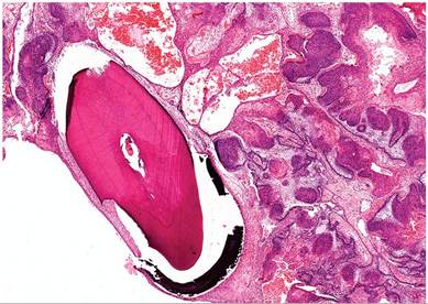
FIG. 6.108. Pseudo-odontoma arising from the elodont incisor tooth root of an aged rabbit. These lesions contain various tooth germ elements, and are often diagnosed as odontomas, but are probably hamartomas.
true neoplasms, but are rather believed to be hamartomas. Thus, the term pseudo-odontoma is the most appropriate descriptor of this syndrome.
Other Neoplasms
Like any species, rabbits develop sporadic cases of neoplasia involving virtually any tissue. Although not as common in commercial and laboratory rabbits because of their relatively younger age, a variety of neoplasms are found in aging pet rabbits. See Heatley and Smith, and Tinkey et al. for more comprehensive reviews on rabbit neoplasia, and von Bomhard et al. for cutaneous neoplasia in rabbits.
BIBLIOGRAPHY FOR NEOPLASMS
General References for Neoplasms
Heatley, J.J. & Smith, A.N. (2004) Spontaneous neoplasms of lago- morphs. Veterinary Clinics of North America Exotic Animal Practice 7:561-577.
Tinkey, P.T., Uthamanthil, R.K., & Weisbroth, S.H. (2012) Rabbit neoplasia. In: The Laboratory Rabbit, Guinea Pig, Hamster and Other Rodents (eds. M.A. Suckow, K.A. Stevensn, & R.P. Wilson), pp. 447-501. Elsevier.
Von Bomhard, W., Goldschmidt, M.H., Shofer, F.S., Perl, L., Rosenthal, K.L., & Mauldin, E.A. (2007) Cutaneous neoplasms in pet rabbits: a retrospective study. Veterinary Pathology 44:579-588.
Uterine Adenocarcinoma
Asakawa, M.G., Goldschmidt, M.H., Une, Y., & Nomura, Y.
(2008) The immunohistochemical evaluation of estrogen receptoralpha and progesterone receptors of normal, hyperplastic, and neoplastic endometrium in 88 pet rabbits. Veterinary Pathology 45:217-225.Green, H.S.N. (1958) Adenocarcinoma of the uterine fundus in the rabbit. Annals of the New York Academy of Science 75:535-542.
Lymphosarcoma
Fox, R.R., Meier, H., Crary, D.D., Meyers, D.D., Norberg, R.F., Sc Laird, C.W. (1970) Lymphosarcoma in the rabbit: genetics and pathology. Journal of the National Cancer Institute 45:719-730.
Gomez, L., Gazquez, A., Roncero, V., Sanchez, C., & Duran, M.E. (2002) Lymphoma in a rabbit: histopathological and immunohistochemical findings. Journal of Small Animal Practice 43:224-226.
Ishikawa, M., Maeda, H., Kondo, H., Shibuya, H., Onuma, M., & Sato, T.A. (2007) A case of lymphoma developing in the rabbit cecum. Journal of Veterinary Medical Science 69:1183-1185.
Kolappaswamy, K., Kriel, E.H., McLeod, C.G., & DeTolla, L.J. (2006) Intermittent inappetence and fur loss in a New Zealand White rabbit. Laboratory Animals (NY) 35:19-20.
Reed, S.D., Shaw, S., & Evans, D.E. (2009) Spinal lymphoma and pulmonary filariasis in a pet rabbit (Oryctolagus cuniculus domes- ticus). Journal of Veterinary Diagnostic Investigation 21:253-256.
Ritter, J.M., von Bomhard, W., Wise, A.G., Maes, R.K., & Kiupel, M. (2012) Cutaneous lymphomas in European pet rabbits. Veterinary Pathology 49:846-851.
Toth, L.A., Olson, G.A., Wilson, E., Rehg, J.E., & Claassen, E. (1990) Lymphocytic leukemia and lymphosarcoma in a rabbit. Journal of the American Veterinary Medical Association 197:627-629.
Volopich, S., Gruber, A., Hassan, J., Hittmair, K.M., Schwenden- wein, I., & Nell, B. (2005) Malignant B-cell lymphoma of the Harder's gland in a rabbit. Veterinary Ophthalmology 8:259-263.
White, S.D., Campbell, T., Logan, A., Meredith, A., Schultheiss, P., Van Winkle, T., Moore, P.F., Naydan, D.K., & Mallon, F. (2000) Lymphoma with cutaneous involvement in three domestic rabbits (Oryctolagus cuniculus). Veterinary Dermatology 11:61-67.
Thymoma
Florizoone, K. (2005) Thymoma-associated exfoliative dermatitis in a rabbit. Veterinary Dermatology 16:281-284.
Vernau, K.M., Grahn, B.H., Clarke-Scott, H.A., & Sullivan, N. (1995) Thymoma in a geriatric rabbit with hypercalcemia and periodic exophthalmos. Journal of the American Veterinary Medical Association 206:820-822.
Wagner, F., Beinecke, A., Fehr, M., Brunkhorst, N., Mischke, R., & Gruber, A.D. (2005) Recurrent bilateral exophthalmos associated with metastatic thymic carcinoma in a pet rabbit. Journal of Small Animal Practice 46:369-370.
Mammary Adenoma and Adenocarcinoma
Baba, N. & Von Haam, E. (1972) Animal model: spontaneous adenocarcinoma in aged rabbits. American Journal of Pathology 68:653-656.
Baum, B. & Hewicker-Trautwein, M. (2015) Classification and epidemiology of mammary tumours in pet rabbits (Oryctolagus cuniculus). Journal of Comparative Pathology 152:291-298.
More on the topic NEOPLASMS:
- Other Neoplasms
- NEOPLASMS
- Nonlymphoid Hematopoietic Neoplasms
- BIBLIOGRAPHY FOR NEOPLASMS
- BIBLIOGRAPHY FOR NEOPLASMS
- Content
- Histiocytic Sarcoma
- Cervical and Perianal Neoplasias
- LEUKEMIA
- The very origins of the laboratory mouse were promulgated by an interest in the genetic basis of cancer, and this interest has accelerated with GEMs.