Histiocytic Sarcoma
Histiocytic sarcomas occur most often in SD rats, but they also have been observed in other strains. The tumors are present primarily in animals over 12 months of age, and there is no obvious sex predisposition.
At necropsy, sarcomas of this type may be present in the liver, lymph nodes, lung, spleen, mediastinum, retro- peritoneum, or subcutaneous tissue. Neoplasms are pale and moderately firm, and they tend to infiltrate and displace normal tissue. Necrotic areas may be scattered in the mass. On microscopic examination, tumors consist of diffuse sheets of neoplastic cells, varying from elongated, pallisading fusiform cells to plump, pleomorphic histiocytic cells. The histiocytic cells have vesicular nuclei, prominent nucleoli, and abundant cytoplasm. Multinucleated giant cells are usually present in tumors with a prominent histiocytic component (Figs. 2.89 and 2.90). Based on electron microscopic and immunohistochemical studies, the histiocytic forms are derived from monocytes or histiocytes, while the origin of the fibrous types remains uncertain. Differential diagnoses include fibrosarcoma, lymphosarcoma, osteosarcoma, and granulomatous inflammatory tissue.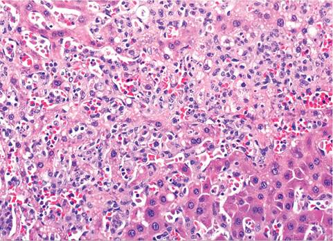
FIG. 2.89. Histiocytic sarcoma infiltrating the liver of an aged rat. Note the indistinct cytoplasmic outlines, anisokaryosis, and pleomorphic appearance of the histiocytic cells that are dissecting through hepatic cords.
More medical literature on Medic.Studio
More on the topic Histiocytic Sarcoma:
-
Infectious diseases -
Internal diseases -
Obstetrics and Gynaecology -
Pediatrics -
Veterinary medicine -
-
Conflictology -
Ecology -
Economy -
Finance -
History -
Law -
Medicine -
Philosophy -
Religious studies -