Lymphoma and Lymphocytic Leukemia
Lymphoma and lymphocytic leukemia are relatively uncommon in most strains of rats. At necropsy, splenomegaly, enlarged lymph nodes (Fig. 2.87), and hepatomegaly are characteristic findings.
On microscopic examination, there is frequently diffuse infiltration of neoplastic lymphocytes into organs such as spleen and liver, with obliteration of the normal architecture. Primary thymic lymphomas have also been described.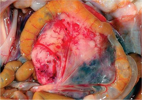
FIG. 2.87. Lymphoma in a rat arising from mesenteric lymphoid tissue. Other than LGL leukemia, lymphoid tumors are rare in rats. (Source: D. Imai, University of California, Davis, CA. Reproduced with permission from D. Imai.)
Cutaneous Lymphoma: Mycosis Fungoides Epidermotropic lymphomas are relatively rare in rats. Clinically, the disease is characterized by the presence of circumscribed erythematous plaques on the skin that may progress to ulceration. Microscopically, there is epidermal hyperplasia with variable ulceration and marked infiltration with neoplastic lymphocytes in the dermis and epidermis. In the epidermis, the infiltrating cells occur singly or in clusters surrounded by a clear halo (Fig. 2.88). Similar changes are present in hair follicles in the region. The infiltrating lymphocytes are medium to large size and react with anti-CD3 antibody, providing confirmation that they are of T-cell origin. In cases documented to date, the neoplastic infiltrates have been confined to the skin.