NEOPLASMS
Hamsters were once commonly used for the experimental induction of tumors by a number of xenogeneic viruses, including adenoviruses, papillomaviruses, and polyomaviruses. Spontaneous tumors are relatively rare among hamsters.
There is a marked variation in the prevalence of neoplasms in different colonies. This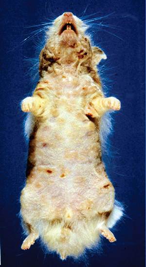
FIG. 3.37. Aged hamster with multifocal raised ulcerated areas on the skin and erythroderma due to epidermotropic lymphoma.
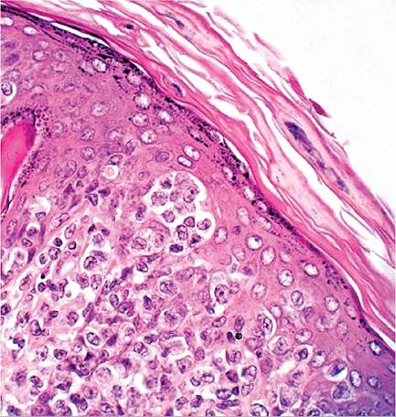
FIG. 3.38. Skin from an aged hamster with epidermotropic lymphoma. Note the infiltrate of poorly differentiated mononuclear cells in the dermis and excavation of the adjacent epidermis.
probably reflects the influence of genetic and environmental conditions. Lymphomas and epithelial tumors associated with hamster polyomavirus (HaPyV) have been discussed previously in the “Viral Infections” section. In addition, spontaneous lymphomas arise in aged hamsters that are not associated with HaPV. They are multicentric, often involving thymus, thoracic lymph nodes, mesenteric lymph nodes, superficial lymph nodes, spleen, liver, and other sites. Cell types are variable. Cutaneous lymphoma resembling mycosis fun- goides has been observed in adult hamsters. Lethargy, anorexia, weight loss, patchy alopecia, and exfoliative erythroderma have been observed in affected animals (Fig. 3.37). Microscopic changes include infiltrates of neoplastic lymphocytes in the dermis, with extension into the epidermis (Fig. 3.38). During the 1960s and 1970s, a contagious reticulum cell sarcoma was known to occur within some laboratory hamster colonies. Tumor cells were transmissible by direct contact and by feeding. Although not reported in recent years, conditions may allow recurrence of this phenomenon.
Among other tumors that occur in this species, the majority are benign, and frequently arise from the endocrine system or alimentary tract. Adrenocortical adenomas are among the most frequently recorded tumors. For additional information on neoplasms, see Pour et al. (1976), Pour et al. (1979), Strandberg (1987), Turusov et al. (1996), Van Hoosier and Trentin (1979), and Barthold (1996).
More on the topic NEOPLASMS:
- BIBLIOGRAPHY FOR NEOPLASMS
- Nonlymphoid Hematopoietic Neoplasms
- Other Neoplasms
- NEOPLASMS
- NEOPLASMS
- NEOPLASMS
- NEOPLASMS
- NEOPLASMS
- Myeloproliferative Neoplasms
- BIBLIOGRAPHY FOR NEOPLASMS
- BIBLIOGRAPHY FOR NEOPLASMS
- Associated Neoplasms Kaposi Sarcoma
- Mesenchymal Neoplasms Rhabdomyosarcomas
- Neurological complications of HIV-1 infection, due either to the immunosuppression (opportunistic infections and neoplasms) or the neurotropism of the virus, are common and add considerably to the morbidity and mortality of the infection
- Neoplasia of the Reproductive System
- BRAIN TUMORS
- Cervical and Perianal Neoplasias
- Cardiac Tamponade
- Bloodstream Infections and Catheter-Related Bloodstream Infections
- Mesothelioma