Mesothelioma
These neoplasms are occasionally encountered in laboratory rats, particularly the F344 strain. Affected rats frequently present with ascites, and at necropsy, there are multiple raised circumscribed yellow to brown nodules present in both the peritoneal and pleural cavities (Figs.
2.95 and 2.96). In most cases, the primary site is the tunica vaginalis of the testes, with subsequent implantation on the serosal surfaces of the peritoneal and pleural cavities. Microscopically, there are diffuse to nodular aggregations of cuboidal to polyhedral cells on serosal surfaces. It is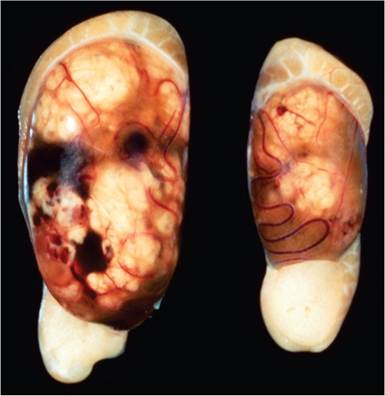
FIG. 2.93. Testes from an aged Fischer 344 male rat, illustrating multiple interstitial (Leydig) cell tumors arising in both testes.
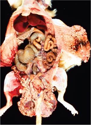
FIG. 2.95. Fischer 344 rat with diffuse mesothelioma involving both the abdominal and thoracic cavities. Note the multiple raised circumscribed polypoid lesions on the serosal surfaces.
Λ*-'∙⅛,÷' r-⅛>':
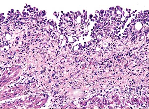
FIG. 2.96. Mesothelioma on the pericardium of a Fischer 344 rat, depicting the papillary growth of neoplastic mesothelial cells on a fibrovascular stromal base.
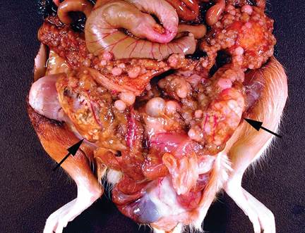
FIG. 2.97. Mesothelioma and interstitial cell tumors (arrows) in an aged rat. Concommitant tumors are common in rats.
fairly common to have simultaneous interstitial cell tumors in cases of mesothelioma (Fig 2.97).