Mesenchymal Neoplasms Rhabdomyosarcomas
Rhabdomyosarcomas are generally uncommon, but arise more frequently in some strains of mice. A strain mice, which carry a mutation in the dysferlin gene which results in development muscular dystrophy, also develop a very high frequency (>70%) of rhabdomyosarcomas at greater than 20 months of age.
Tumors are pleomorphic and arise from the axial and proximal appendicular skeleton. It has been proposed that regeneration of skeletal muscle due to the dysferlin mutation has a promoting effect upon tumor development, since muscles that are most affected by muscular dystrophy are most prone to neoplasia. Aside from A strain mice, BALB mice are more prone to development of rhabdomyosarcomas than other strains of mice. Quadriceps muscle is the most frequent site for these tumors in BALB mice (Fig. 1.141).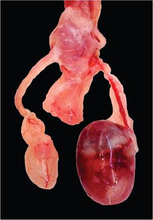
FIG. 1.139. Enlarged testis (right) in a 129 mouse due to growth of a teratoma. (Source: A. Haertel, Davis, CA. Reproduced with permission from A. Haertel.)
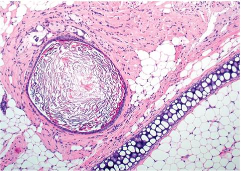
FIG. 1.140. Testicular teratoma from a mouse. This field illustrates the presence of well-differentiated cartilage, adipose tissue, muscle, an epithelial lined cyst filled with keratin, and adjacent sebaceous glands.
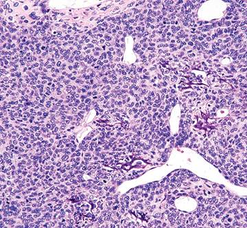
FIG. 1.142. Femoral osteosarcoma. The tumor is highly cellular with sparse osteoid formation.
Osteomas and Osteosarcomas
Primary bone tumors are relatively rare in nonmanipulated strains of laboratory mice. Exceptions are outbred OF-1 and CF-1 mice, which develop benign, frequently multicentric osteomas in up to 30% of the mice. A relatively high incidence (7%) of osteosarcomas has been noted to develop in NOD and NOD-derived substrains of mice (Fig.
1.142). Tumors arise primarily in the appendicular skeleton, and particularly the femur. Bone tumors are also relatively common in Trp53 null heterozygous mice. Rare osteosarcomas arise sporadically in other strains of mice, and have been noted to arise from spinal vertebrae, sternebrae, or long bones. Possible sites for metastatic spread include lung, liver, spleen, and kidney. The lumbosacral region appears to be the most common site for primary tumors arising from the vertebral column. Tumors at this site frequently impinge on the spinal cord, resulting in a clinical presentation of posterior paresis or paralysis. In advanced cases, changes in the adjacent spinal cord are consistent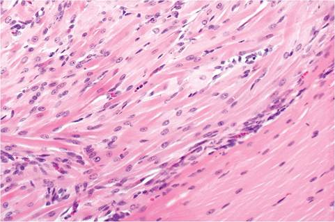
FIG. 1.141. Rhabdomyosarcoma arising from the quadriceps muscle (lower right) of a BALB mouse. The tumor is composed of disorganized neoplastic myocytes (strap cells).
with Wallerian degeneration. Natural infection of nude mice with polyoma virus has been associated with vertebral bone tumors and posterior paresis.
Other Mesenchymal Tumors
Mesenchymal tumors can be readily induced by carcinogens and viruses, such as Moloney murine sarcoma virus. Soft tissue sarcomas are common in some GEMs, especially Trp53 homozygous and heterozygous null mice, with tumor prevalence partially determined by mouse strain background. Sarcomas can be readily induced in nearly 80% of Trp53 heterozygous mice following subcutaneous implantation of transponders or plastic foreign bodies.
Endocrine Neoplasms
Pituitary gland adenomas are relatively common in B6 and Swiss mice (Fig. 1.143). FVB/N mice are particularly prone to development of these tumors. Most pituitary
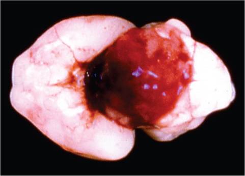
FIG. 1.143. Pituitary adenoma in a Swiss mouse. These tumors grow by expansion and are typically red because of sinusoids filled with blood.
adenomas produce prolactin and arise more frequently in females. Tumor cell growth patterns may be solid, sinusoidal, or cystic, with the latter two types appearing quite bloody. These tumors grow expansively, with compression of the overlying brain. Carcinomas are less common and are more anaplastic and invasive. Adrenocortical adenomas, pheochromocytomas, pancreatic islet tumors, follicular cell adenomas of the thyroid gland, and other endocrine types of tumor occur sporadically in laboratory mice and are usually represented in “lists” of tumors arising in aged mice.
More on the topic Mesenchymal Neoplasms Rhabdomyosarcomas:
- BIBLIOGRAPHY FOR NEOPLASMS
- Nonlymphoid Hematopoietic Neoplasms
- Other Neoplasms
- NEOPLASMS
- NEOPLASMS
- NEOPLASMS
- NEOPLASMS
- NEOPLASMS
- NEOPLASMS
- Myeloproliferative Neoplasms