Hepatocellular Neoplasia
Spontaneous hepatic neoplasms in the mouse include hepatocellular adenomas and carcinomas, hepatoblastoma, cholangioma, cholangiocarcinoma, cholan- giohepatocellular (mixed) carcinoma, hemangioma, hemangiosarcoma, and histiocytic sarcoma.
Rarely, Ito cell tumors may also be encountered. The most common hepatocellular tumors are adenomas and carcinomas, which arise more frequently in aged males than females. Some strains of mice, such as A and DBA, are especially prone to hepatocellular tumors (Fig. 1.132), and infection with Helicobacter spp. has been associated with an earlier onset and higher prevalence among A strain mice. Foci of cellular alteration, including clear cell foci, basophilic cell foci, and eosinophilic cell foci, are generally considered to be antecedent to hepatocellular neoplasia. Primary hepatic tumors readily occur in mice treated with a variety of hepatocarcinogens.Hepatocellular tumors may vary from single to multiple, circumscribed, raised, moderately firm, gray to tan nodules to large, poorly delineated, pale to dark red fleshy masses. Hepatocellular tumors are composed of two major histological types: trabecular (Fig. 1.133) or solid (Fig. 1.134), but other patterns may arise, including adenoid. Cellular morphology may be well differentiated to poorly differentiated, and the degree of differentiation does not predict metastatic potential. Well-differentiated hepatocellular adenomas may be difficult to distinguish from adjacent tissue. Most hepatocellular tumors are well circumscribed and not encapsulated, but some
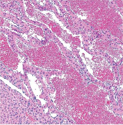
FIG. 1.133. Hepatocellular carcinoma, trabecular type. The neoplasm consists of cords of neoplastic hepatocytes growing in a trabecular pattern.
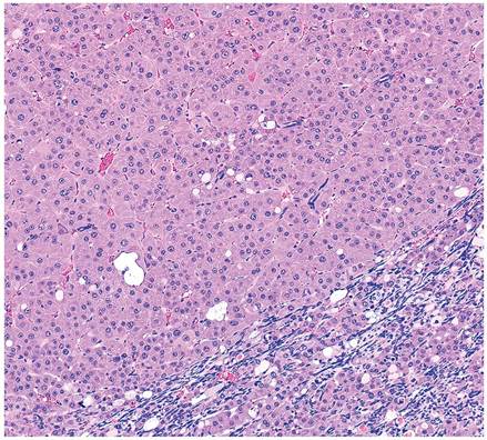
FIG. 1.134. Hepatocellular carcinoma, solid type. The adjacent liver parenchyma (lower right) is compressed by the tumor.
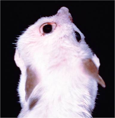
FIG. 1.135. Mouse with retrobulbar Harderian gland adenocarcinoma resulting in facial distortion and periocular porphyrin staining.
may be locally infiltrative. Anisokaryosis, karyomegaly, and cytomegaly are frequently prominent. A rare variant is hepatoblastoma, which has characteristic organoid structures arranged around vascular channels or forming rows and rosettes. Cholangiomas and cholangiocarcino- mas are also rare. Hemangiomas and hemangiosarcomas are somewhat more common and may arise as primary liver neoplasms. The liver is frequently involved in multisystemic lymphoreticular neoplasms and may occasionally be the site of metastatic dissemination of other tumor types.