Lawsonia intracellularis Infection
Along with other species (particularly hamsters and rabbits), rats are susceptible to infection with L. intra- cellularis. Natural infection of laboratory Wistar rats has been reported, in which multiple rats were described that developed adenocarcinomas of the ascending colon.
With Warthin-Starry staining and electron microscopy, lesions contained typical intracytoplasmic bacteria within the apical cytoplasm of hyperplastic enterocytes. At the time, the bacteria were considered to be Campylobacter-like organisms, and C. jejuni-like bacteria were isolated from affected rats, contributing to the assumption of etiology. In retrospect, the intracytoplasmic bacteria had pathognomonic features of L. intracellularis. Lesions resembled the invasive hyperplastic crypt epithelium and histiocytic inflammation in hamsters infected with L. intracellularis, with formation of multiple cystic granulomatous subserosal nodules and occasional extension into mesenteric lymph nodes. In all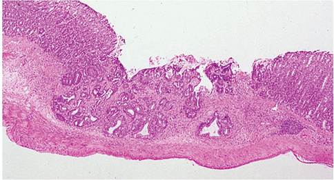
FIG. 2.35. Helicobacter-associated colitis in an athymic rat. The colonic mucosa is hyperplastic, with focal crypt atypia and crypt herniation. (Source: J.M. Ward, Montgomery Village, MD. Reproduced with permission from J.M. Ward.)
probability, the lesions were not neoplastic, but the epithelial invasiveness typical of this disease has led to that conclusion in hamsters as well.
Salmonella enterica Infection
Salmonella enterica infects and produces disease in a wide variety of animals, including humans. For this reason, it can be introduced to laboratory animal populations in a number of ways. During the early years of the 20th century, Salmonella infections represented an important infectious disease in laboratory rodents. However, with improved sanitation, health-monitoring methods, and feeding practices, the disease now rarely occurs in laboratory animal colonies.
During 1895-1910, serotype Enteritidis was used as a rodenticide to control populations of wild rats in Europe and the United States. Enthusiasm waned when the public health implications became apparent. Of the multitude of serotypes of S. enterica capable of causing disease in the laboratory rat, serotypes Enteritidis and Typhimurium have been most frequently implicated.Pathology
Clinical signs include depression, ruffled hair coat, porphyrin crusting around the eyes, hunched posture, weight loss, and variations in the nature of the feces from softer, lighter, formed feces to fluid feces. Sub- clinical infections without discernible lesions are frequent. In clinically affected rats, the ileum and cecum are often distended with liquid contents and flecks of blood, and there may be thickening of the gut wall in affected areas. Focal ulcerations may be present in the mucosa of the cecum and ileum. Splenomegaly frequently occurs. On microscopic examination, lesions in the ileum and cecum are characterized by hyperplasia of crypt epithelial cells, edema of the lamina propria, and leukocytic infiltration with focal ulceration. There is hyperplasia of the mesenteric lymph nodes, spleen, and Peyer's patches, with focal necrosis and leukocytic infiltration. In acute cases, lesions in other viscera are consistent with a Gram-negative septicemia, with focal embolization within spleen, liver, and lymph nodes. Emboli consist of bacteria, fibrinous exudate, and cellular debris. In the spleen, focal granulomas, fibrinous exudation, and focal necrosis are typical lesions present in the red pulp. Sinusoidal congestion and focal coagulation necrosis are frequent findings in the liver.
Diagnosis
Isolation and identification of the organism from animals with lesions or from inapparent carriers are necessary to confirm the diagnosis of S. enterica infection. Salmonellae are intermittently present in the intestine, especially in carrier animals. Repeated fecal samplings may be required in order to detect inapparent carrier animals. At necropsy, mesenteric lymph nodes are a tissue of choice, as they tend to yield Salmonella in rats with negative fecal culture. Differential diagnoses include Tyzzer's disease, pseudomoniasis, C. jejuni infection, Enterococcus enteritis, rotaviral enteritis, cryptosporidiosis, and management-related problems due to failure to provide feed or water.