Lawsonia intracellularis Infection: Proliferative EnterocolitisZHistiocytic Enteritis
Lawsonia intracellularis is an obligate intracellular bacterium that does not grow in cell-free medium, although some success has been achieved with growth in cell cultures. Colonization of enterocytes by this organism has been identified in several species, including mice, rats, hamsters, guinea pigs, rabbits, and rhesus macaques as well as numerous other species.
It is a significant pathogen in pigs and an emerging pathogen in horses. Proliferative and histiocytic lesions of the small intestine are the typical changes associated with L. intracellularis infections. For a number of years, the causative agent was called an intracellular Campylobacter-like organism. Based upon 16S rRNA sequencing, there appears to be little genetic variation among organisms from different host species, but interspecies susceptibility varies with different isolates. Transmission is likely to be by ingestion of fecal material, and experimental transmission has been achieved in this manner. There is no evidence that the organism infects tissue other than enteric mucosa. Location of lesions (gut segment) varies with species. In the rabbit, the jejunum and proximal ileum are typically affected. The fact that the location of lesions is typical for each host species suggests that intestinal microenvironment or host receptors may be critical to pathogenesis. The means of cellular invasion is not known, but in vitro studies indicate that it involves receptor-ligand mechanisms. Regardless of host species, L. intracellularis organisms grow to large numbers within the apical cytoplasm of enterocytes.Pathology
In acute infections, diarrhea with mortality occurs in suckling, weanling, and young adult domestic rabbits. In animals necropsied during the acute disease, semifluid mucinous contents are frequently present, particularly in the colon and rectum. Animals with more chronic lesions have thickened opaque loops of small intestine (Fig.
6.37). The mucosal surfaces are rugose in character. Microscopically, mucosal lesions vary from suppurative and erosive to primarily proliferative in nature. Lesions of the erosive type range from focal denuding to segmental loss of enterocytes, with polymorphonuclear cell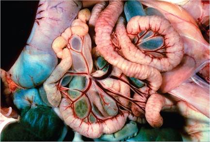
FIG. 6.37. Histiocytic enteritis in a laboratory rabbit associated with Lawsonia Intracellularis infection. Note the marked thickening and rugose appearance of the serosal surface of the small intestine.
infiltration in the underlying areas. Proliferative lesions are characterized by multifocal to diffuse hyperplasia of enterocytes lining crypts and villi, with mononuclear cell infiltration. Histiocytes with abundant granular cytoplasm, and occasionally multinucleated giant cells, are often prominent in the lamina propria (Fig. 6.38). Lacteals are often dilated. Silver- and PAS-stained sections of affected mucosa reveal typical intracytoplasmic clusters of small bacteria in the apical cytoplasm of the crypt-villus column (Fig. 6.39). Histiocytes within the lamina propria have PAS-positive granular material in their cytoplasm. Electron microscopy reveals typical organisms in enterocytes, and histiocytes contain
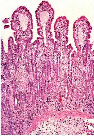
FIG. 6.38. Jejunum from a laboratory rabbit with histiocytic enteritis due to Lawsonia intracellularis infection. Villi are shortened, lacteals are dilated, crypts are hyperplastic, and the lamina propria and submucosa are infiltrated with histiocytes and focal accumulations of lymphocytes. Source: © R J Hampson
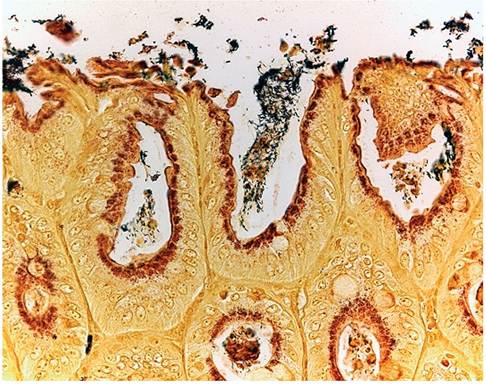
FIG. 6.39. Intestinal mucosa from a rabbit infected with Lawsonia intracellularis. Note the dense argyrophilic populations of bacteria within the apical cytoplasm of enterocytes (Warthin-Starry stain).
degenerating bacterial debris. Marginal subclinical infections are common in enzootically infected rabbit colonies, with identification of lesions as incidental findings at necropsy. Coinfection with enteropathogenic E. coli has been documented in rabbits.
Diagnosis
The identification of the typical proliferative mucosal lesions and the demonstration of the intracellular organisms within the apices of enterocytes with the appropriate silver stains are confirmatory. The organism can be grown in cell culture. Other procedures described include the demonstration of the organism in fecal samples using immunomagnetic beads or PCR, the identification of bacterial surface antigen in tissue sections by immunohistochemistry, and the detection of antibodies to L. intracellularis in sera.