Listeria monocytogenes Infection:
Listeriosis
Listeriosis was originally described in 1926 by Murray et al. in young rabbits and guinea pigs. At that time, the agent was named Bacterium monocytogenes. Listeria monocytogenes is a small Gram-positive, nonspore-forming rod with zoonotic potential.
Recently, analysis of rabbit meat in Europe revealed it to be a significant source of food-borne Listeria. Listeriosis in rabbits is characterized by abortions and sudden deaths, particularly in does during advanced pregnancy.Epizootiology and Pathogenesis
Listeria is a soil-borne organism. In sporadic outbreaks of the disease, the source of the organism is frequently attributed to contamination of feed or water. Inapparent carriers and shedders may also occur. Listeria monocytogenes has a specific predilection for the gravid uterus in advanced pregnancy. Adult, nonpregnant does and bucks are usually resistant to the infection. Following oral or conjunctival inoculation of females in advanced pregnancy, abortions, stillbirths, and mortality in the dam usually occur. Pregnancy may be interrupted as early as 24 hours postinoculation. However, inoculation of females by the intravaginal, oral, or conjunctival route either prior to mating or early in pregnancy failed to produce disease. The organism can cross the placental barrier in advanced pregnancy. Uterine infections may persist postkindling and may serve as the source of infection for the next pregnancy. On the other hand, young rabbits may shed Listeria as an inapparent infection for several weeks postkindling.
Pathology
Deaths typically occur in does in advanced pregnancy. Straw-colored fluid is frequently present in the peritoneal cavity, occasionally with fibrinous exudate and ecchymo- ses on the serosal surface of the uterus. Disseminated pale, miliary foci of necrosis in the liver, edema of regional lymph nodes, splenomegaly, and visceral congestion are the usual macroscopic findings.
The uterus may contain relatively intact, near-term kits (Fig. 6.40) or fetuses in various stages of decomposition or mummification. In acute cases, the placenta may be edematous and hemorrhagic, but in an infection of longer duration, the placenta is usually thickened, friable, and dull dirty gray, with an irregular surface. Characteristic microscopic changes seen in adult cases of listeriosis include focal hepatitis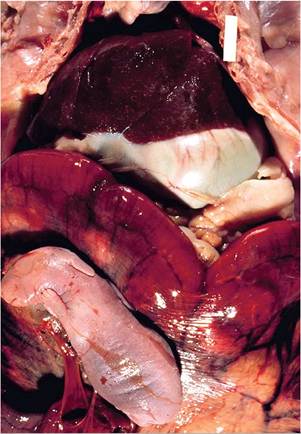
FIG. 6.40. New Zealand White doe that died near-term with acute
listeriosis. There are pinpoint-size foci of hepatitis. The externalized kit (lower left) is intact, with no evidence of maceration (Courtesy R.J. Hampson.)
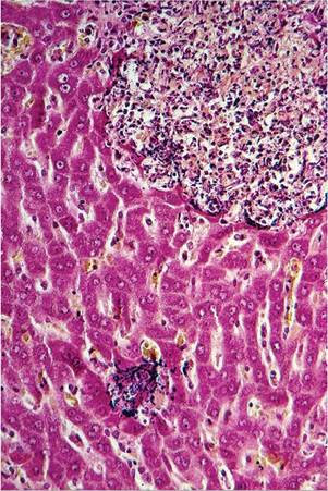
FIG. 6.41. Focal hepatitis from a case of listeriosis in a pregnant doe. Note colonies of Gram-positive bacteria (Brown and Brenn stain).
(Fig. 6.41). There may be focal inflammatory lesions in the adrenal cortices, congestion and thromboses in the splenic sinusoids and blood vessels, and acute necrotizing to chronic suppurative metritis and placentitis. In rabbits that die with the acute form of the disease, large numbers of Gram-positive bacilli are usually visible, particularly in the placenta. Focal hepatitis and occasionally meningitis have been observed in newborn kits that succumb within a few days of birth. Kits that survive may subsequently develop systemic listeriosis, or may present with stunting and meningoencephalitis.
Diagnosis
In acute cases of listeriosis, the organism can usually be readily recovered from the uterine wall, placenta, and fetuses. Blood, liver, and spleen are other likely sources of the organism at necropsy. Recovery of the organism by culture is considered to be a much more satisfactory method to confirm the diagnosis than are serological techniques. Isolation is enhanced when tissues to be cultured are held at 4°C for several days before inoculating culture plates.
PCR methods have also been used for rapid diagnosis. Differential diagnoses include diseases causing disseminated foci of hepatic necrosis such as Tyzzer's disease, tularemia, and salmonellosis. In perinatal deaths seen in does with acute pasteurellosis and metritis, there may be acute necrotizing uterine lesions, but liver lesions are normally absent.Moraxella bovis Infection
A rabbit that was housed in close proximity to cattle was described with suppurative metritis, pleuritis, pneumonia, and focal hepatic necrosis. Lesions were confirmed to be positive for Moraxella bovis.
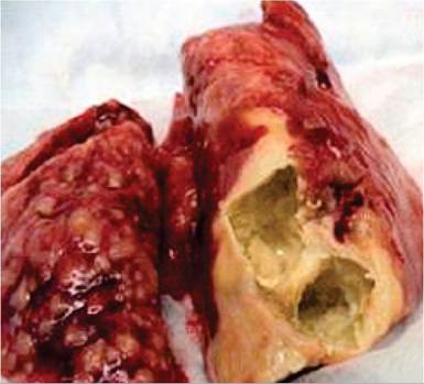
FIG. 6.42. Pulmonary tuberculosis in a rabbit infected with Mycobacterium tuberculosis. Rabbits develop cavitary pulmonary lesions with frequent dissemination to other organs. (Source: Nedeltchev et al, 2009. Reproduced with permission from American Society for Microbiology.)
More on the topic Listeria monocytogenes Infection::
- CHAPTER 35 LISTERIA INFECTIONS
- Picornavirus Infection: Mouse Encephalomyelitis Virus Infection
- Arenavirus Infection: Lymphocytic Choriomeningitis Virus Infection
- Arterivirus Infection: Lactate Dehydrogenase-Elevating Virus Infection
- Coronavirus Infection: Mouse Hepatitis Virus Infection
- Streptococcus pneumoniae Infection: Pneumococcal or Diplococcal Infection
- INFLAMMATION IN HIV-1 INFECTION
- Blastomyces dermatitidis Infection
- Mycobacterium Avium Complex Infection
- Characteristics of Infection and Disease
- Astrovirus Infection
- COVID-19 INFECTION
- IN HIV-1 INFECTION