Lungs
The right and left Iurujs lie within the thoracic cavity on either side of the double layer of connective tissue known as the mediastinum (Figs 8.7 and 8.8). Each lung consists of the air passages (only the most proximal parts of the bronchi are outside of the lung), blood vessels and surrounding connective tissue, all enclosed within a membrane called the pulmonary pleura (Fig.
8.5A).'I hc lungs are divided into well delined lobes by deep fissures (Fig. 8.9). The left lung is divided into three lobes; the right lung has four lobes. The lobes are referred to as the crania!or apical, middle or cardiac, and caudal or diaphragmatic. The fourth lobe of the right lung is the accessory lobe; this is small and lies on the medial surface of the caudal lobe of the right lung.
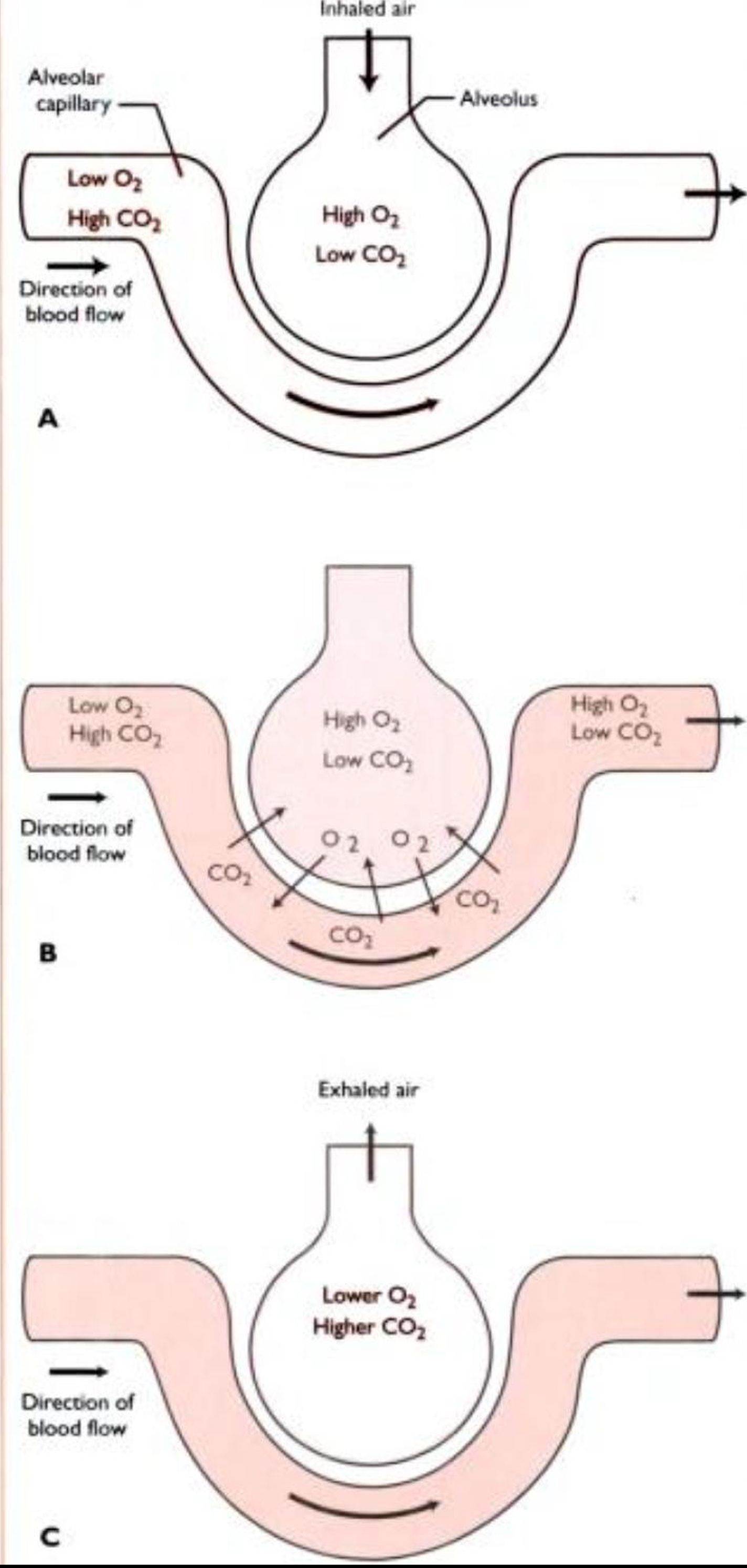
Fig. 8.6 The exchange of gases wιthn the alvco∣∣ A l∏χ>rabon the air
contains high IeveE of oxygen and low levels of carbon dioxide. Bkxxj cntcmg the.Ilvcolar capillary contains taw levels of Oxygen and high
levels of cartoon d∣θχ∣de B Gis exchange occurs oxygen diffuses from ⅛r ∣n the alvcotas. where its level ∣s high, into blood m the.alveolar capillary, where its level β taw. Carbon doχ∣de does the reverse, diffusing from alveolar capillar into alveolus C Expiration: exhaled a∣r contains less oxygen and more carbon dioxide than are present in room air Next breath bongs m a fresh supply of high oxygen air (Repnnted from Cltmcal Anatomy and Physology for Vetennary Technicians. T Colville and ]M Bassett, p 233. Gopynght 2002. with permission from Elsevei Science.)