Myoepitheliomas
Myoepitheliomas arise infrequently in most strains of mice but are relatively more common in some strains, such as BALB/c and BALB/cBy mice, especially females.
They most frequently arise from submaxillary and parotid salivary glands but may also be associated with mammary, preputial, and Harderian glands.
These tumors can become very large, with cystic chambers containing mucinoid fluid (Fig. 1.137). Microscopically, tumors are composed of large, pleomorphic spindle cells with epithelial and mesenchymal features (Fig. 1.138). Cystic areas form as a result of necrosis. Metastasis to the lung may occur with large tumors. A curious feature is concomitant myeloid hyperplasia of bone marrow and spleen, apparently related to a secretory product of the tumor.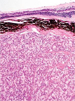
FIG. 1.136. Harderian gland adenocarcinoma depicted in Figure 1.135. The neoplasm consists of poorly differentiated fusiform to cuboidal epithelial cells, with compression of the adjacent sclera and retina.
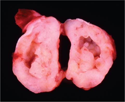
FIG. 1.137. Parotid salivary gland myoepithelioma of a mouse. The tumor has been bisected to demonstrate the cystic center that contained necrotic cellular debris and mucinous material.
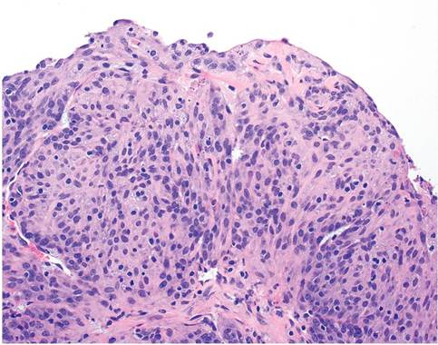
FIG. 1.138. Myoepithelioma, demonstrating the morphology of the epithelioid/spindle-shaped neoplastic cells lining the cystic center.