Otitis media is a common disease process that often goes unrecognized in most veterinary practices.
The fact that otitis media is present in more than half of all canine patients with chronic otitis externa should stimulate a reformulation of the diagnostic process when faced with these cases.
Just the common history that the patient has been treated repeatedly for ear infections should alert the veterinarian to think about otitis media as a possibility.The diagnosis of otitis media in dogs can be quite difficult to make, owing to the long, bent, funnel-shaped conformation of the dog’s ear canal, which makes visualizing the tympanic membrane (TM) difficult. In addition, many patients with otitis media have an intact TM, giving the clinician the impression that there is nothing wrong in the middle ear. Most canine patients with otitis media also have a chronic otitis externa with pathologic changes to the ear canal that cause stenosis, making visual examination of the TM impossible. It is often theorized that otitis media is an extension of otitis externa that was either not treated, improperly treated, or resistant to treatment. The result is significant damage, resulting in porosity to the eardrum over time.
Otitis media in cats most often results as a sequela to respiratory disease, so a history of sneezing, ocular discharge, and/or nasal discharge may provide a clue. Some cats with otitis media also have a visible polyp in the ear canal after the ear is cleaned of the dried exudates and mucus. Many feline otitis media cases have a dark, dried, crumbly exudate in the ear canal that mimics an ear mite infestation. The diagnosis of otitis media in cats may be easier to determine with the otoscope because of their relatively short ear canals.
Otitis media should be considered when the clinician is presented with a patient showing any neurologic disease affecting the head, including vestibular disease, Horner’s syndrome, or facial nerve damage.
By definition, otitis media refers to the extension of an inflammatory disease into the middle ear cavity. This may or may not be infectious. The inflammatory reaction of the mucous membrane lining the tympanic bulla is different than the reaction of the skin of the external ear, so the symptomatology and treatment of otitis media are different from those for otitis externa. The mucous membrane lining the tympanic bulla reacts to foreign substances, including infectious organisms, hair, cells, cerumen from the external ear canal, chemicals, and pharmaceuticals used in the external ear canal. It produces a purulent exudate and increases its secretion of protective mucus from activated goblet cells.
If the eardrum has a hole in it during active otitis media, copious mucoid exudate is often seen along the floor of the horizontal canal. Although this material is usually in liquid form, the mucus and pus may be inspissated and dry. Mucus is not produced anywhere along the external ear, but oozes from the tympanic bulla into the horizontal canal through any rent in the TM. The presence of mucus indicates a hole in the eardrum; precautions should be taken not to introduce ototoxic substances into the ear canal.
Otitis media in dogs is much more prevalent than previously thought. In dogs, secondary otitis media occurs in approximately 16% of acute otitis externa cases and as many as 50% to 80% of chronic otitis externa cases.1,2 Many cases of otitis media are well hidden from visual detection by the significant exudates present in the ear canal and the severe pathologic changes that have occurred in the ear canal as a result
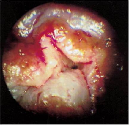
Figure 14-1
Stenotic ear. Extensive fibrosis in the horizontal ear canal prevents visual examination of the TM.
of chronic otitis externa. These changes include stenosis, fibrosis, tumors, polyps, epithelial hyperplasia, and glandular hyperplasia (Figure 14-1). Chronic changes in the external ear canal prevent adequate visualization beyond these blockages, so determining the integrity of the eardrum is not always possible.
Some dogs with otitis media have intact eardrums but also have significant bacterial and yeast populations that can be isolated from the middle ear.2 These dogs may have had a ruptured eardrum that healed, trapping bacteria and yeast in the tympanic bulla. Therefore the presence of an eardrum does not rule out otitis media. Healed eardrums trap infectious organisms in the middle ear, and suppurative otitis media results (Figure 14-2). Secretions and exudates are trapped behind the healed eardrum, causing it to bulge outward under pressure, which in turn causes severe pain. Myringotomy may be necessary to investigate the contents of the bulla in suspected cases of otitis media where the eardrum is intact.
More on the topic Otitis media is a common disease process that often goes unrecognized in most veterinary practices.:
- Otitis media is a common disease process that often goes unrecognized in most veterinary practices.
- One of the most common ailments of dogs seen in a veterinary practice is ear disease.
- Etiolog y of Ceruminous Otitis
- Treatment of Otitis Media
- Otitis interna, or infection of the inner ear, is a relatively common disorder in the dog.
- Primary Otitis Media in Cats
- Environmental and Conformational Causes of Ceruminous Otitis
- Otitis externa is a common malady, occurring in 15% to 20% of dogs and 5% to 7% of cats seen in veterinary practice.
- Most cases of acute otitis externa are pruritic.
- The Use of Corticosteroids in Otitis Media