Polyarteritis
Inflammatory lesions of small and medium-sized arteries are common in many strains of laboratory mice. The character of the lesions varies and may include fibrinoid degeneration and necrosis of the tunica media and inflammation associated with neutrophilic and/or mononuclear leukocytes.
There may be thickening and fibrosis of the vascular wall. The distribution of affected vessels is quite variable but most often involves arteries of the tongue, head, pancreas, heart, kidneys, mesentery, urinary bladder, uterus, testes, and gastrointestinal tract, among others. Lesions tend to be segmental, feature different stages of acute to chronic inflammation, and involve multiple vessels. The etiology of polyarteritis is not known, but immune complexes have been demonstrated within affected vessels. It is common in mice that are prone to autoimmune disease, including MRL and NZB mice. Polyarteritis is usually an incidental finding, but it can be associated with segmental infarction of the kidneys with scarring. It may also have vestibular manifestations when vessels of the head are involved (see “Vestibular Syndrome”).Atrial Thrombosis and Heart Failure
Thrombosis of the auricle is a frequent finding in mice, which may manifest as left- or right-sided heart failure (Fig. 1.114). The left auricle is most frequently affected. Atrial thrombosis typically involves organizing thrombi in the auricle, but the process may extend into the ventricles, cardiac vessels, and pulmonary veins. This syndrome is typically precipitated by multisystemic amyloidosis, but it is also relatively common in BALB/ c mice, which are not prone to amyloidosis. Left heart failure due to atrial thrombosis is the most common cause of noninfectious dyspnea in mice.
Perivascular Lymphoid Infiltrates
Mild to severe infiltrates of lymphoid cells may arise in the adventitia of pulmonary vessels, with extension into adjacent alveolar septa. This is invariably in response to antigenic stimuli, such as a prior virus infection. They should not be present in pathogen-free mice. They also frequently appear in older mice with perivascular mononuclear cell infiltrates in salivary glands, kidneys, and other organs. These infiltrates seem to be antecedent to lymphoproliferative disorders.
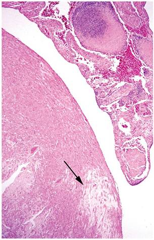
FIG. 1.114. Heart of a mouse with auricular thrombosis. Note the organizing thrombus within the auricular lumen and the patch of amyloid infiltration in the myocardium (arrow). Auricular thrombosis is often associated with renal disease, including amyloidosis.