DIPHTHERIA
Diphtheria is a life-threatening but vaccine preventable disease of childhood, characterized by acute severe inflammation of upper respiratory epithelium with membrane formation and marked toxemia.
Incidence has declined in recent years, due to expanding immunization.However, India still contributes to more than half of the cases of diphtheria globally and recent years have seen its resurgence in some geographical regions.
Epidemiology: Diphtheria is caused by three toxigenic strains (gravis, intermedius and mitis) of C. diphtheria (Klebs-Loffler bacillus)-gram positive non-invasive bacilli, almost exclusively found at respiratory mucous membranes of clinical cases or asymptomatic carriers.
Infection is usually transmitted as droplet infection or via contaminated fomites/ dust, though direct contact with infected secretions is a rare source of infection.
Infectivity period lasts for ~2-4 weeks from the onset of disease, except in chronic carriers (gt;1 month). A case or carrier is considered as non-infective, only when at least two throat cultures at 24-hour interval are negative.
Diphtheria is most common in preschool children (lt;5 years), during autumn and winter season and unvaccinated children living in overcrowded environment. Pathogenesis: Major virulence of the organism lies in its ability to produce an exotoxin, responsible for local necrosis and systemic complications. On entry, C. diphtheriae proliferates superficially at the site of inoculation (usually respiratory epithelium) to produce dense necrotic material consisting of organisms, epithelial cells, fibrin, leukocytes and erythrocytes and an adherent grayish-white pseudomembrane that bleeds on attempted removal.
Clinical features: After incubation period of 2-5 days, these cases present with
• Constitutional symptoms, e.g. acute onset of moderate fever, typical toxic look and altered mental state, e.g.
confusion or irritability. Circulatory collapse is not uncommon.• Local respiratory tract symptoms, depending on the site of lesion, as follows:
± Pharyngotonsillar diphtheria, the commonest type, presents with sore throat, dysphagia, submandibular swelling (bull-neck) and a grayish- white dirty membrane on soft palate, tonsils and pharyngeal wall that bleeds on removal (Fig. 10.1).
± Laryngotracheal diphtheria, the most serious type, presents with acute hoarseness of voice, croupy cough, stridor and progressive respiratory distress due to airway obstruction by membrane and tissue edema.
± Nasal diphtheria, the mildest form, is characterized by serosanguineous or blood-mixed nasal discharge with membrane formation and shallow ulcers at nasal mucosa and external nares.
• Non-respiratory disease is uncommon, presenting as cutaneous diphtheria (punched-out, tender ulcers with membrane), conjunctival diphtheria (purulent/ ulcerative conjunctivitis), aural diphtheria (otitis externa) and genital diphtheria (purulent or ulcerative vulvovaginitis).
Complications may be due to: (a) airway obstruction by membrane or edema, (b) aspiration of infected material, (c) exotoxin production, and (d) abnormal immune response, e.g. Guillain-Barre syndrome (Table 10.10).
Airway obstruction is the immediate cause of mortality in most cases of diphtheria, though important late complications include:
• Toxic myocarditis, usually during 2nd week of illness presenting with tachycardia, muffled heart sounds, CCF and arrhythmia. Seen in 10-25% cases, it is responsible for ~ 50-60% of late deaths in diphtheria.
• Cranial nerve palsies usually present as palatal palsy in 2nd-3rd weeks with nasal twang, regurgitation of feeds and aspiration, or occular palsies in 3rd-5th weeks with loss of accommodation (earliest sign), blurred vision and squint.
• Post-diphtheritic peripheral neuropathy may be sensory, motor or combined, involves one or more limbs and usually develop in 2nd-3rd weeks.
• Guillain-Barre syndrome is a rare late complication after 6-8 weeks and indicates immunological insult.
Diagnosis of diphtheria depends on:
• Clinical presence of tough dirty-white membrane in throat that bleeds on removal,
• Direct microscopy of throat/nasal swabs with Albert's stain to demonstrate the characteristic club-shaped
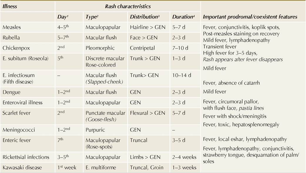
Fig. 10.1: Diphtheria (oropharyngeal membrane).
TABLE 10.10: Complications of diphtheria
Respiratory:
• Airway obstruction gt; respiratory failure
• Secondary bacterial pneumonia
• Atelectasis
• Late: Bronchiectasis, lung abscess
Cardiac:
• Toxic myocarditis and CCF
• Endocarditis (extremely rare)
Neuropathies:
• Cranial: Palatal or ocular palsy
• Peripheral: Monoparesis
• Guillain-Barre syndrome
Renal:
• Proteinuria (secondary nephrotic syndrome)
• Acute renal failure
Others: Septicemia, hepatitis, arthritis
bacilli with metachromatic staining, arranged in a chinese-letter pattern. Fluorescent antibody staining is more sensitive for this purpose.
• Throat/nasal swab culture on Loffler or tellurite media to confirm the diagnosis and differentiate from diphtheroids—non-pathogenic oropharyngeal commensals.
• A rapid ELISA test, PCR test or modified Elk test may be used to detect presence of diphtheria toxin.
• Ancillary investigations, e.g. ECG, urine examination, etc. help to exclude complications.
D/D depends on the site of lesion:
• D/D nasal diphtheria: Other causes of serosanguineous or blood-mixed discharge, e.g. (a) viral rhinitis, (b) foreign body and (c) snuffles (congenital syphilis).
• D/D pharyngotonsillar diphtheria: Other causes of throat membrane, e.g. (a) streptococcal membranous tonsillitis, (b) viral membranous tonsillitis, e.g. infectious mononucleosis, (c) oropharyngeal candidiasis, (d) post tonsillectomy membrane.
• D/D laryngeal diphtheria: Other causes of acute croup and stridor (see Table 16.8).
Management: Considering high mortality, morbidity and infectivity, all suspected cases of diphtheria need hospitalization. Important steps in management include:
• Strict isolation till at least two consecutive nose/ throat swabs at 24-hour interval are negative.
• Specific anti-toxin therapy to neutralize free circulating toxin, given as early as possible.
Currently, only equine diphtheria antitoxin (ADS) is available. Doses are empirical, depending on the site of membrane and duration of illness, i.e. 40,00060,000 IU for nasopharyngeal lesions, 20,000-40,000 IU for pharyngeal or laryngeal lesions of lt;48 hours and 80,000-1,20,000 IUfor severe airway obstruction or longer illness. It is given as slow IV infusion over 30-60 minutes after a subcutaneous test dose (0.02 ml) to detect hypersensitivity.
• Antibiotic therapy to stop further toxin production and eliminate organisms. Drug of choice is Crystalline penicillin (IV 1-1.5 lac U/kg/d q6hr) or Erythromycin (PO 40-50 mg/kg/d q6hr), given for 14 days or till two successive cultures are negative. However, antibiotic therapy is not a substitute for antitoxin therapy.
• Management of complications: Immediate tracheostomy is indicated in severe respiratory distress. Even in mild cases, hospitalization with complete bed rest is indicated for 2 weeks—to monitor for late complications, to reduce risk of myocarditis and to prevent spread of infection.
Prevention: Diphtheria is preventable by:
• Active immunization of all children with three primary doses at 6, 10 and 14 weeks of age and two boosters at 18 months and 5 years, followed by Td every 10 years from 10 years onwards (see Ch 9.2.1). IAP also recommends a single dose of Tdap vaccine after 7 years, followed by Td at 17-18 years.
• Isolation and treatment of symptomatic cases as well as asymptomatic carriers. All carriers must be treated with PO Erythromycin (40-50 mg/kg/d q6hr) for 10-14 days or till two throat-cultures are negative. ADS is not required in asymptomatic cases, even if culture is positive.
• Management of close contacts: While all close contacts should be cultured from nasopharynx to detect asymptomatic carriers, chemoprophylaxis with PO Erythromycin (40-50 mg/kg/day for 7-10 days) or a single IM Benzathine Penicillin 6 Lac IU (12 Lac IU above 30 kg) is necessary in all close contacts.
All immunized contacts should also be given a booster dose of DPT/DT, if the last dose was received gt;2 years ago. Unimmunized contacts must be started on primary immunization schedule.
10.6
More on the topic DIPHTHERIA:
- DIPHTHERIA
- PASSIVE IMMUNIZATION
- STRABISMUS (SQUINT)
- OTHER CALF DISEASES
- ACUTE CROUP
- DEMOGRAPHIC AND SOCIAL ASPECTS OF EUROPEAN EXPANSION
- NATIONAL IMMUNIZATION SCHEDULE
- Infectious Agents
- MISSION INDRADHANUSH
- INFECTIOUS CAUSES