Electromyography
Motor Unit Configuration and Amplitude
Amplitudes of motor unit action potentials (MUAPs) are lower in infants, with amplitudes ranging from 150 microvolts to approximately 2,000 microvolts.
Generally, motor unit action potentials more than7.4
Normal Sensory Conduction Velocities (m/sec)
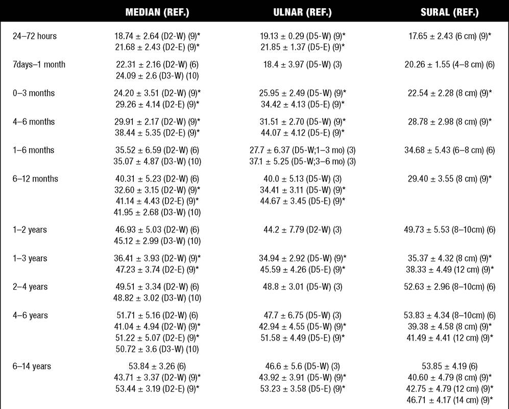
Data are presented as means ± SD
*Velocities based on peak latencies for Cai and Zhang (9); others based on onset latencies
D-W = Finger to wrist using ring electrodes with orthodromic stimulation
D-E = Finger to elbow using ring electrodes with orthodromic stimulation
D2 = Index finger for median; D3 = middle finger for median; D5 = fifth finger for ulnar
Sural nerve studies use antidromic with recording electrodes behind the lateral malleolus with stimulus delivered at 6 cm to 14 cm above the malleolus as specified.
1,000 microvolts in 0- to 3-year-old children are rare (24,25). In infants, motor unit action potentials are usually biphasic or triphasic.
Motor Unit Duration
Infantile motor unit action potentials are often shorter in duration. DeCarmo (24) found newborn infants to exhibit durations 17% to 26% shorter than those seen in adults. Durations of motor unit action potentials are often shorter than 5 milliseconds in infants.
Motor Unit Recruitment
In very young infants and children, it is difficult to assess strength of voluntary contraction and determine when the interference pattern is full. In general, as strength of voluntary contraction increases, there is an increase in motor unit action potentials recruited. However, the recruitment pattern in infants may be disordered and chaotic. As with adults, the recruitment frequency, defined as the firing rate of a MUAP when a different MUAP first appears, with gradually increasing strength of voluntary contraction, is helpful
7.5
Normal Sensory Nerve Action Potential (SNAP) Amplitudes in Children (μV)
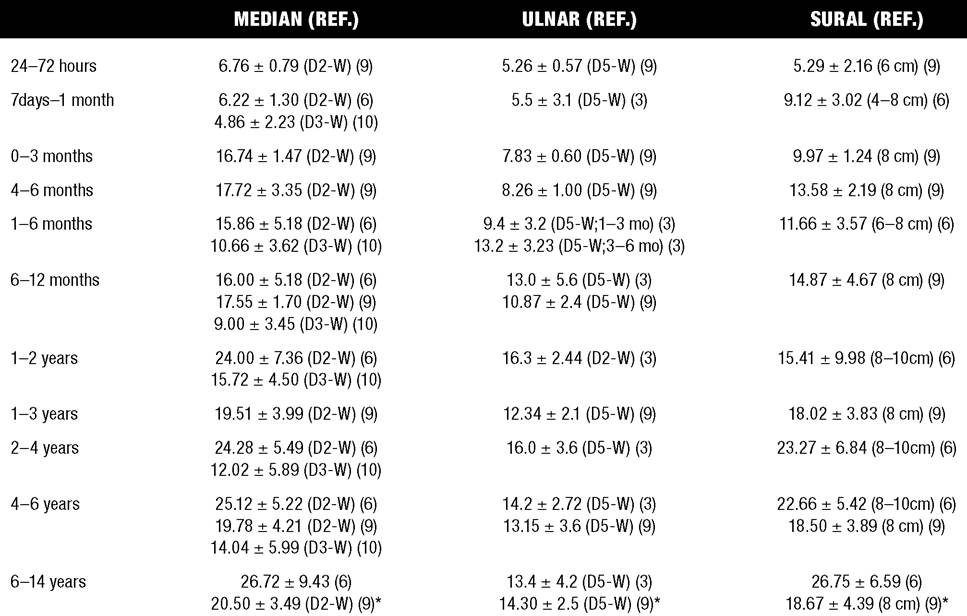
Data are presented as means ± SD
Amplitudes are determined peak-to-peak from positive-to-negative peak of the SNAP
D-W = Finger to wrist using ring electrodes with orthodromic stimulation
D2 = Index finger for median; D3 = middle finger for median; D5 = fifth finger for ulnar
Sural nerve studies used antidromic stimulation with recording electrodes behind the lateral malleolus with stimulus delivered at 6 cm to 14 cm above the malleolus as specified.
in differentiating a myopathic process (lower recruitment frequency values) from a neuropathic process (higher recruitment frequencies after greater than 20-25 Hz). An example of neuropathic recruitment is shown in Figure 7.1.
More on the topic Electromyography:
- Electrodiagnostic Studies
- TECHNICAL FACTORS OF NEEDLE ELECTROMYOGRAPHY
- Electrodes
- Repetitive Nerve Stimulation Studies
- Stimulating Electrodes
- Volume Conduction
- Entrapment Mononeuropathies in Children
- Prefac
- Recording Electrodes
- NEUROMUSCULAR DISORDERS