PERICARDITIS
Pericarditis denotes infective or non-infective inflammation of the pericardium, which may or may not be associated with excess fluid collection in pericardial sac (pericardial effusion), secondary compression of heart with compromised filling (constrictive pericarditis) or severe limitation of cardiac output (cardiac temponade).
Normal pericardial sac contains ~10-15 ml of serous fluid as shock absorber and lubricant.Etiopathogenesis: Tuberculosis is the commonest cause of exudative pericardial effusion in India, followed by bacterial infections, e.g. Staph. aureus (pyopericardium). Infection may reach pericardium by direct spread from neighboring foci, e.g. pneumonia, empyema, osteomyelitis of ribs, etc. or via hematogenous spread from a distant foci, e.g. meningitis.
Transient pericardial effusion is common in viral infections (? hypersensitivity), while collagen disorders are usually associated with large effusions and polyserositis in older children (Table 17.35).
Pathophysiology: Depending on the cause, pericardial fluid may be serous, fibrinous, purulent or hemorrhagic. Except in small serous effusions, rising intra-pericardial pressure due to accumulated fluid affects cardiac functions and may cause cardiac tamponade, characterized by progressive limitation of the ventricular diastolic filling and reduction in stroke volume. In most cases,
TABLE 17.35: Causes of pericarditis
• Infective
- Bacterial: Staphylococci, streptococci, pneumococci
- Tuberculosis
- Viral: Coxsackie B, EBV, adenovirus
- Parasitic: E. histolytica, hydatid disease
- Fungal: Histoplasmosis, actinomycosis
• Inflammatory
- Rheumatic fever
- Collagen disorders: Rheumatoid arthritis, SLE
• Hematological: Leukemia, bleeding diathesis
• Traumatic chest injuries (hemopericardium)
• Iatrogenic: Post-pericardiotomy, radiotherapy
• Others: Uremia, hypothyroidism
• Idiopathic
the effusion resolves spontaneously or on treatment.
However in some cases (specially tuberculosis), gradual organization and fibrous thickening of pericardium may lead to obliteration of the pericardial space, i.e. constrictive pericarditis.Clinical manifestations depend upon the amount, type and rapidity of fluid collection. Small effusions may be completely asymptomatic.
Common presenting symptoms include—(a) dull or stabbing pre-cordial or left-shoulder pain due to diaphragmatic/pleural irritation, which aggravates on supine position and relieves on sitting and leaning forward, (b) cough, dyspnea, dysphagia and hiccough due to bronchial, esophageal or phrenic nerve compression, (c) constitutional symptoms, e.g. fever, weight loss, etc. or (d) features of primary disease. Physical findings depend on whether the pericarditis is associated with effusion or cardiac tamponade.
• Pericarditis with minimum effusion presents with Pericardial friction rub — a scratching, grating, high- pitched sound, best heard with the diaphragm of stethoscope applied firmly at the lower left sterna border in sitting and leaning-forward position. Friction rub disappears with increasing fluid collection.
• Moderate effusion without cardiac tamponade is characterized by—(a) quiet pericardium with ill-defined apical impulse, (b) cardiomegaly on percussion with cardiac dullness extending beyond the apex beat, (c) muffling of the heart sounds, and
(d) peripheral signs of poor diastolic filling, e.g. tachycardia, raised JVP, congestive hepatomegaly and basal lung crepitations.
• Cardiac tamponade or constrictive pericarditis presents with severe peripheral signs due to poor diastolic filling, e.g. (a) low volume pulse with pulsus paradoxus, (b) distended neck veins with Kussmaul's sign-paradoxical increase in JVP during inspiration, (c) tender, congestive hepatomegaly, and (d) hypotension due to compromised cardiac output.
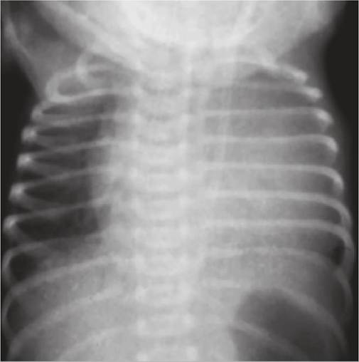
Fig.
17.25: Pericardial effusion.Note: Narrow cardiac pedicle and oligemic lung fields
Diagnosis: USG is the best method to detect very small pericardial effusions, while in others, diagnosis is supported by:
• Chest X-ray showing cardiomegaly with narrow pedicle due to compression of great vessels, i.e. Pear-shaped heart and oligemic lung fields (d/d CCF with usually plethoric lung fields). However, heart size is reduced in constrictive pericarditis (Fig. 17.25).
• ECG with-(a) low-voltage QRS complexes due to dampening effect of pericardial fluid, (b) elevated ST segment due to myocardial irritation, and (c) inverted T waves due to myocardial inflammation.
• USG is diagnostic, revealing presence of an echo-free space between the epicardium and pericardium. Presence of both, anterior as well as posterior effusions, indicates large effusions.
• Echocardiography is useful to detect impending cardiac tamponade/constrictive pericarditis, indicated by—
(a) flattening of septal motion, (b) collapse of RA in late diastole, and (c) collapse or indentation of RV free wall (specially outflow tract) in diastole.
• Pericardiocentesis with biochemical analysis (exudate vs. transudate) and culture of aspirated fluid is necessary for etiological diagnosis. On gross appearance, pericardial fluid is—(i) Xanthochromic in tubercular pericarditis, (ii) Thick purulent in bacterial infections, e.g. Staph. aureus, (iii) Serofibrinous in viral or rheumatic pericarditis and connective tissue disorders, and (iv) hemorrhagic in malignancy, trauma and bleeding disorders.
• Relevant investigations for etiological diagnosis, e.g. tuberculin test, ASO titers, etc.
Management of pericardial effusion depends on primary etiology and includes:
• Specific treatment, e.g. antibiotics in pyopericardium; antitubercular drugs plus steroids in tubercular effusions; and steroids in rheumatic fever or collagen disorders.
• General supportive measures, e.g. bed rest, oxygen, analgesics, and nutritional support. Decongestive therapy with diuretics and digoxin should be avoided due to risk of further compromise in cardiac output.
• Surgical interventions, i.e. pericardiocentesis is indicated in pyopericardium or cardiac tamponade. Constrictive pericarditis requires decortication of pericardium with radical pericardiectomy.
17.11
More on the topic PERICARDITIS:
- PERICARDITIS
- Constrictive Pericarditis
- Pathology of the Pericardium
- Mycobacterium Tuberculosis
- Pericardial Effusion and Tamponade
- Streptococcus pneumoniae Infection: Pneumococcal or Diplococcal Infection
- Chemotherapy
- Lymphoma
- Streptococcus and Enterococcus Infections
- PNEUMOCOCCAL INFECTIONS