Common ear ailments of dogs and cats can be seen in up to 50% of patients in areas of humid climates during some or all of the patient’s life.
Dogs have a moderately higher incidence of ear disease than cats.
Treatment of the variety of inflammatory, infectious, neoplastic, and chronic connective tissue changes that can be observed in small animal patients is often very difficult, unrewarding, and frustrating.
This is due to the restricted size of the external auditory meatus and tympanic bulla. The small size of these areas is often further restricted in the presence of disease due to excessive inflammation of the dermis and underlying tissues. Recurrences are common. This may be due to a failure to resolve the underlying pathology or incomplete treatment (resection) of the problem. Repeated treatments, assessments, and an extended duration of therapy are not uncommon. This often causes increased client concern and dissatisfaction.Implementation of laser energy for the effective treatment of a variety of conditions can simplify both the therapeutic protocol and the recovery of the patient. Video otoscopy offers the opportunity to enhance the diagnostic ability of the veterinarian. Because tissues are visualized on a video monitor under magnification, the condition of the ear can be more accurately assessed. In combination with a surgical laser, many diseases of the ear canal and bulla can be treated in an aggressive, yet minimally invasive manner. The advent of advanced otoscopic equipment coupled with CO2 and/or diode-wavelength laser energy can significantly improve the final therapeutic outcome for many diseases of the ear. This method of therapy can lead to shorter recovery times with less discomfort for the pet.
Video otoscopic equipment has greatly enhanced the veterinarian’s ability to evaluate and treat diseases of the ear canal and middle ear (bulla). A variety of systems are available. These systems can be broadly categorized as diagnostic or therapeutic systems.
The major difference is the presence of an operating channel in the therapeutic systems. For the purposes of this discussion, only therapeutic systems are considered.Therapeutic endoscopic telescopes are usually either the 0 degree (directly forward) or 30 degree (oblique view). Video otoscopes are 0 degree. They are usually larger (around 4.75 mm [because of the presence of an operating channel]), have a larger field of view, and are more durable and less costly than other rigid endoscopes. The single working channel of the video otoscope allows access for suction, biopsy, irrigation, and laser devices. The 2-mm channel accepts instrumentation up to 5 Fr, or about 1.8 mm. The biopsy channel can provide for simultaneous irrigation, suction, and treatment with the use of a two- or three-port connector, as shown in Figure 19-1.
A 2.7- ? 180-mm, 0-degree, or 30-degree rigid endoscopic telescope with a 14-Fr operating sheath can also be used in these procedures. The smaller overall diameter, longer length, and separate irrigation and operating channels make this endoscope an excellent choice for interventional otoscopy (Figure 19-2). The 30-degree endoscope is particularly useful in laser surgery and treating lesions of the middle ear. The 30-degree offset allows for visualization of the laser delivery system or surgical instrumentation as it exits the operating channel (Figure 19-3). This allows for more precise alignment of the instrument and decreases the possibility of iatrogenic damage.
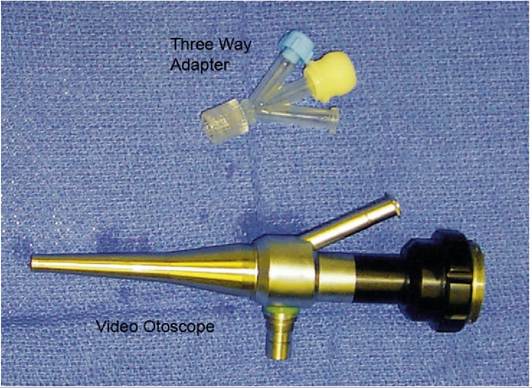
Figure 19-1
Video otoscope and three-way adapter.
Because of the higher cost and increased fragility of this equipment, it should not be used in conscious patients.
Lasers can both extend and enhance the therapeutic options for the treatment of aural disease. Two types of lasers are useful in the treatment of auricular pathology. They are the CO2 laser (10,600-nm wavelength) and the diode laser (either 810- or 980-nm wavelength).
It is beyond the scope of this chapter to discuss fully the physics involved with each of these wavelengths. The practitioner is strongly encouraged to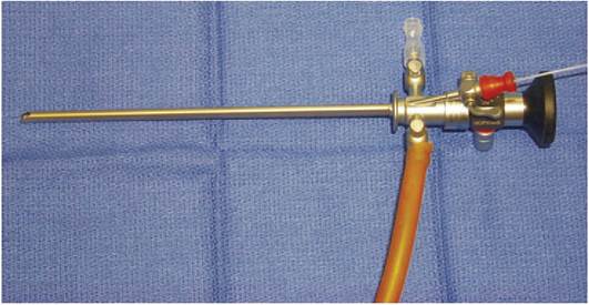
Figure 19-2
Rigid endoscope prepared for suction, irrigation, and laser surgery.
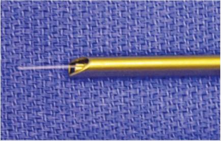
Figure 19-3
Treatment end of 30-degree rigid endoscope.
review and understand the concepts of power density and tissue interactions before using this technology. By combining lasers and video otoscopy, a wider range of problems can be corrected in a minimally invasive fashion. This often leads to a better outcome. Lasers can prove extremely useful in the following:
• Aural hematoma repair
• Removal of masses in the aural canal
• Controlling hemorrhage during video otoscopy
• Ear canal resection and ablation
• Removal of masses in and around the pinna
• Treatment of chronic otitis
• Myringotomies
• Cosmetic and therapeutic otoplasty (ear trims, correction of abnormal ear carriage, and treatment of neoplasia)
In general lasers can be used in three different modalities:
1. Coagulation. In this mode lasers are used to provide hemostasis. This is accomplished by providing laser energy at a lower power, usually with a longer exposure time. By using the laser in this manner, tissue contraction rather than vaporization is achieved. When the collagen in the end of a bleeding vessel is contracted, hemostasis is achieved.
2. Incision. By providing relatively high wattage with a small focal point, high power densities are achieved. This results in incision of the target tissue. This is particularly useful in situations where a mass is pedunculated and the base of the mass can be visualized. Depending on the power density, more or less hemostasis can be attained.
3. Ablation. Laser energy can also be applied to the surface of structures within the ear. In this mode, relatively high energy for moderate exposure times is introduced, and the surface of the targeted structure is vaporized. Through continuous appropriate application of energy, tumors can be removed from all parts of the aural canal and the middle ear.
More on the topic Common ear ailments of dogs and cats can be seen in up to 50% of patients in areas of humid climates during some or all of the patient’s life.:
- One of the most common ailments of dogs seen in a veterinary practice is ear disease.
- Otitis externa is a common malady, occurring in 15% to 20% of dogs and 5% to 7% of cats seen in veterinary practice.
- Selecting the appropriateimaging method, correctly applying the technique selected, and accurately interpreting the examination are the key steps in imaging ear disorders in dogs and cats.
- Most skin diseases may affect the pinna in dogs and cats, but other parts of the body can also be involved.
- Reference ranges for haematology and serum chemistry values for adult cats and dogs
- Otitis interna, or infection of the inner ear, is a relatively common disorder in the dog.
- Careful examination of a clean, dry ear canal in a dog or cat with otitis externa may reveal many conditions that affect the ear canal.
- Primary Otitis Media in Cats
- Proximity to oceans influences regional climates
- The lungs are commonly affected in patients infected with HIV, with over 60% of patients having at least one respiratory episode during the course of their disease.