LAMENESS DUE TO LEG DISORDERS
One of my professors at veterinary school used to say that even if you thought an animal was lame in its head, you should always examine its foot, and I think this is an excellent piece of advice to pass on.
Leg injuries do occur, however, and as you are moving the cow up to the crush to examine her foot, watch the way she walks: if the whole leg is stiff, or being carried, or if it is hanging completely limp, then you may well be dealing with a leg injury.Figure 9.23 shows the names of the bones and joints in the front and hind legs. The correct technical terms will be used throughout this chapter, so be prepared to keep referring back to this diagram. The common leg, pelvis and muscle disorders causing lameness are listed in the following:
Pelvic injuries
knocked down pin bone
split H bone
dislocation of the pelvis (Chapter 5)
Leg and spine injuries
dislocated hip
fractures
spinal abscess and osteomyelitis
Joint problems
arthritis (hip and stifle)
joint ill (Chapter 2) capped knees and hocks cellulitis
copper deficiency (Chapter 12) and rickets (Chapter 12)
Muscle, nerve and tendon injuries obturator and peroneal nerve paralysis (Chapter 5) radial nerve paralysis spastic paresis
(string halt, Elsoe heel) muscle necrosis
(white muscle - Chapter 3) muscle tearing
(downer cow, Chapter 5) rupture of the stifle ligaments
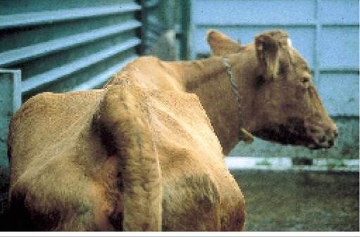
Plate 9.59. Fracture of the wing of the pelvis. Although it looks peculiar, it causes few problems.
rupture of the gastrocnemious tendon (Chapter 5) contracted tendons
overstretched tendons (Chapter 3) popliteal abscess (Chapter 10)
For those conditions which have been covered elsewhere in this book, a page reference has been given and no further mention will be made in the following text.
Calving injuries and the handling of downer cows are discussed in Chapter 5, which should be read in conjunction with this section.Knocked Down Pin Bone (Fracture of the Wing of the Pelvis)
The pin bone is the front wing of the pelvis (Figure 9.23 and Plate 5.7). It can be broken by cows pushing through doorways and other narrow entrances. The cow in Plate 9.59 looks peculiar with one side much lower than the other, but the condition rarely causes any lameness and if the skin is not broken, no treatment is necessary. She will continue to lead a normal productive life. However, if the skin splits and infection enters, as in Plate 9.60, the damage can be very slow to heal. This is especially the case if the bone becomes infected. Removal of all broken fragments of bone and thoroughly cleaning the wound help healing.
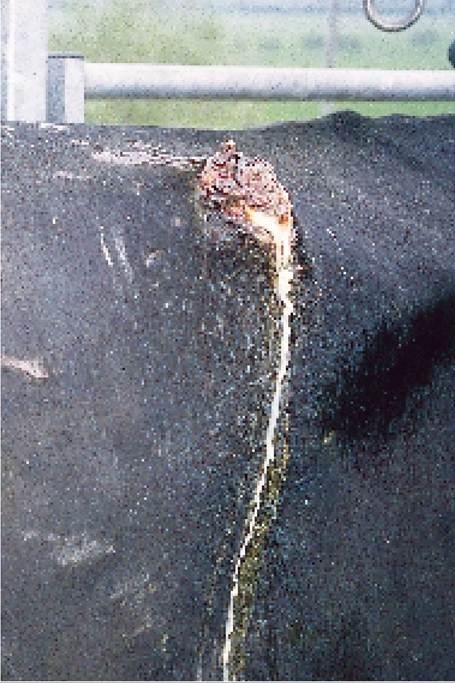
Plate 9.60. Infection of the wing of the pelvis. In this
instance it may take several months for the skin to grow
back.
Split H Bones
The H bone (sometimes written as ‘aitch’ bone) is the name given to the pelvis and so a cow which has ‘split her Hs’ has a broken pelvis. It occurs as a result of a cow ‘doing the splits’, either because she lost her grip on slippery concrete, or because of an injury when she was bulling, or perhaps following obturator nerve paralysis at calving (see Chapter 5).
Dislocated Hip
The normal position of the hip joint is shown in Figure 9.23. Dislocation (sometimes called luxation or subluxation) means that the ball of the upper end of the femur has been forced out of its socket in the pelvis. The head of the femur then pushes forwards, and the ball normally rests on the edge of the pelvis, at the point marked X in Figure 9.23, although occasionally it moves into other positions. It occurs as the result of a severe sprain or twisting of the leg and it is especially common in cows which have been bulling and have fallen on slippery concrete while trying to mount other cows.
This is exactly what happened to the cow in Plate 9.61 and if you look carefully you can see the dislocated hip on her left side, producing a swelling under the skin. I find that the best way to appreciate this is to stand behind the cow with one hand over each hip joint and then let her walk slowly forwards.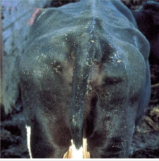
Plate 9.61. Dislocated hip. Aswelling can be seen on the left side of the pelvis.
Very little movement is felt in the normal hip, whereas the dislocated end of the femur will force your hand out and slightly forwards as the cow tries to take weight on the affected leg.
If treatment is to be successful, it must be carried out soon after the injury, before the socket gets filled with blood and the joint becomes too loose. Your vet will sedate the cow and cast her onto her side; then he will extend the affected leg with ropes and pulleys as he tries to push the ball back into the socket. I have had a few successful cases, but many do not respond, or the hip dislocates again as soon as the cow stands up. This probably occurs when the ligaments holding the joint in place have also been totally ruptured. Affected cows may milk on for a while, but if they are already well past peak lactation, it may be better to sell them immediately, before excessive weight loss occurs.
Fractures
Broken legs occur most commonly as a result of cows falling on slippery concrete, again often associated with oestrus. Younger calves may be stood on by cows or get their legs caught in gates. A fracture is diagnosed by moving the leg around, feeling for abnormal movement and listening for the grating sound of bone against bone. It always surprises me that this seems to elicit relatively little pain response from the animal, although weightbearing will probably be zero.
In older animals fracture of the femur is most common and in the majority of cases the cow is recumbent and unable to move.
Treatment of adult fractures is hopeless. In calves, the lower leg is more commonly affected and if a plaster cast or even splints and elastoplast are applied, most will recover well. Fractures through the growth plate of the bone (the epiphyseal plate E) as in Plate 9.62 are the exception to this. A proportion of these fail to heal, or heal very slowly.Spinal Abscess and Osteomyelitis
Spinal abscesses, collapsed vertebrae and general spinal inflammation (osteomyelitis) are all difficult conditions to diagnose. The clinical signs will depend on the position of the lesion in the spine and the tissues
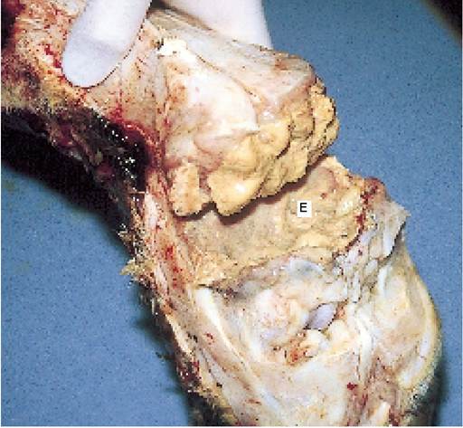
Plate 9.62. Fractures through the epiphyseal plate (E) (growth point of the bone) can be much slower to heal.
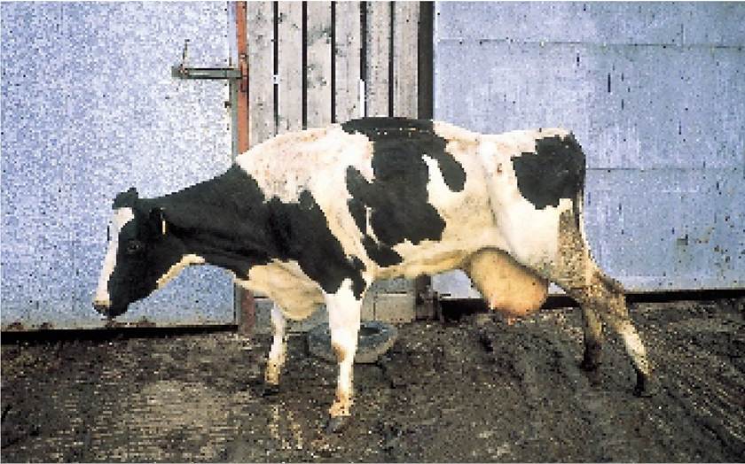
Plate 9.63. Osteomyelitis of the spine. This cow walked with extreme difficulty. Aspinal abscess was found on post-mortem.
involved. Some cows walk very slowly and stiffly, with an arched back (Plate 9.63), others may lose the function of their hind legs, while one cow I dealt with who had an abscess in her cervical (neck) spine was unable to bend her neck and had to kneel down to graze!
Arthritis and Stifle Ligament Rupture
The word means inflammation of the joint. The inflammation could be caused by degeneration due to age, by an infection (for examplejoint ill), by pedal arthritis (page 297) or by excessive movement within the joint. The latter occasionally occurs in the stifle joint (Figure 9.23) of adult cows, which is held together by ligaments. If the ligaments rupture, the two bone surfaces rub across each other and this leads to thickening and new bone formation. The condition is difficult to diagnose in the early stages: the cow has a low-grade lameness and no cause can be found. Later the hard, bony enlargement of one stifle joint becomes obvious.
Rupture of the stifle ligament is a common injury in dogs.A cow with arthritis will have little spicules of jagged bone (see Plate 9.55) protruding from the joint surface and you can imagine the pain caused as the surfaces rub across one another, especially with the weight of the cow pressing on them. Arthritis is most common in older cows, especially in winter. There is no long-term cure, but your vet may be able to suggest anti-inflammatory drugs which will reduce the pain and inflammation in the joint. Moving the cow out of cubicles and into loose housing where it is easier to get up and down will also help.
Capped Knees and Hocks
Soft, fluctuating and painless fluid swellings over the front of the knee and at the side of the hock (Plate 10.25) are quite common, especially in cubicle-housed cows. Plate 9.64 shows an extreme example. The swelling is caused by continual bruising leading to excessive fluid production in the bursa, which is the name given to a type of shock absorber on the outside of the joint. The lesion is not painful and in the majority of cases it is best left alone. Most will slowly disappear after turnout in the spring or after moving the cow into a straw yard.
Sometimes you may wish to drain off the excess fluid. To do this, clip the hair over the centre of the swelling, clean off the area very thoroughly, then insert a sterile needle. A light straw-coloured or sometimes reddish- brown liquid will flow out through the needle. Great care is needed, however, because of the risk of introducing infection and creating an abscess. If the swelling is large and gets damaged it may develop into an abscess and then discharge on its own. This will need flushing out with water and antiseptic ointment infused into the hole to keep it open and promote drainage.
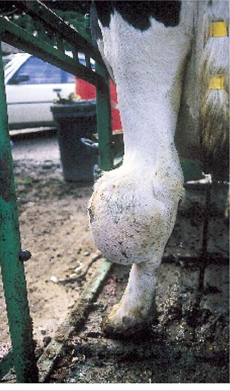
Plate 9.64. A capped hock (hock bursitis) is caused by continual trauma, usually the result of lying on inadequately bedded cubicles.
Cellulitis (Infected Knees and Hocks)
This condition is also seen in cubicle- housed animals, and it is due to infection penetrating through the skin or even into the bursa over the joint. Rather than forming a localised swelling, which we would call an abscess, the infection tracks up and down the leg and causes a more generalised enlargement. This is known as cellulitis and is clearly shown in the right leg of the cow in Plate 9.65. The affected animal will be holding its leg in pain, and there will be some rise in temperature. Treatment consists of giving antibiotics to eliminate the infection and anti-inflammatory drugs to reduce the pain and swelling. In severe cases this may have to be continued for a week or more.
Both capped knees and infections are caused by the same factors; that is, poor housing. Cubicle beds which are rough and have inadequate bedding, or where there is an excessively large or sharp lip at the rear, all predispose to bruising. In some cubicles, the design is such that the hock is knocked on a sharp edge of a wooden division when the cow stands up. This leads to the type of
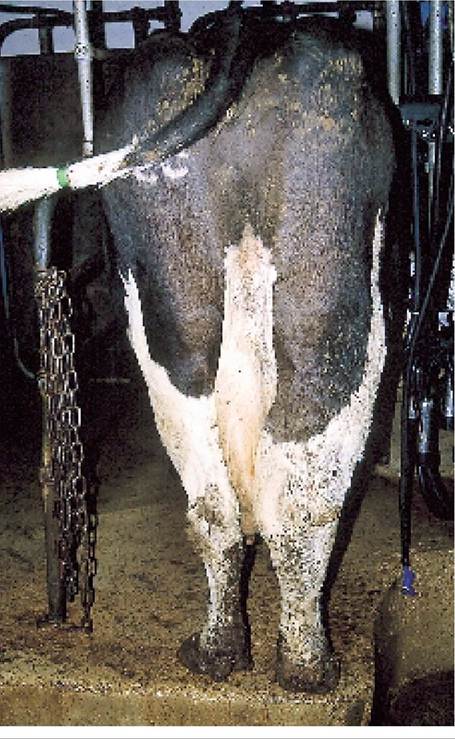
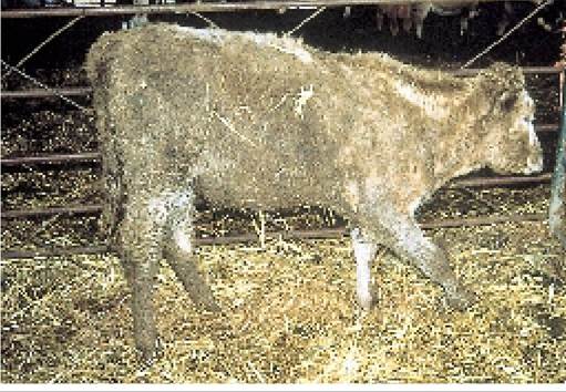
Plate 9.66. Animals with radial paralysis are unable to extend the front leg for weightbearing.
Plate 9.65. Cellulitis is a diffuse infection of the leg tissues. It can make the cow quite ill as well as lame.
injury shown in Plate 9.64. Cows which are lame from other causes also have difficulty getting up, and capped knees or infected hocks may develop secondary to the primary lameness.
Radial Nerve Paralysis
The radial nerve runs from the spine across the chest and into the front leg. Its function is to contract the extensor muscles, thereby extending the leg forwards and stiffening it for weightbearing. An animal with radial paralysis (Plate 9.66) is unable to extend its front leg and cannot bear any weight on it. There is no pain, just the discomfort of finding it difficult to move around. Many animals eventually learn to throw the leg forward at the shoulder and are then able to achieve a degree of weightbearing. The Charolais heifer shown in Plate 9.66 eventually recovered, but took four or five months to do so. Damage to the radial nerve occurs most commonly as a result of the leg being pulled away from the side of the chest, for example getting it caught in a gate or dismounting from a bulling cow.
Spastic Paresis (String Halt, Elsoe Heel)
This is an inherited condition which leads to spasm of the gastrocnemious muscle, and it is most commonly seen in calves aged three to nine months. The leg goes very stiff, is extended backwards (Plate 9.67) and cannot be used for walking. The condition can be corrected surgically by cutting through either the nerve or the gastrocnemious tendon, just above the hock. If the tendon is cut, the leg initially collapses to the ground, like rupture of the gastrocnemious tendon in cows (Plate 5.34), but over a period of two or three months it will return to the upright position.
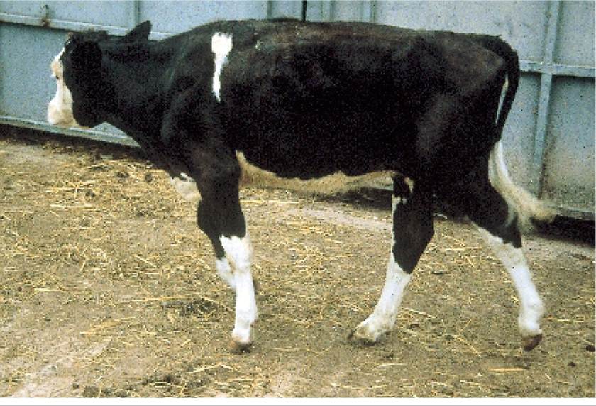
Plate 9.67. Spastic paresis is a nerve disorder resulting in continual spasm of one or sometimes both hind legs.
Contracted Tendons
A proportion of calves are unable to stand at birth because their front legs are buckled over. Figure 9.23 shows the normal position for a front leg and Plate 9.68 shows a calf which cannot straighten its fetlock joint because the flexor tendons running up the back of the leg are too short. The majority of calves slowly improve over two to three weeks and you can help them by providing plenty of room for movement and by lifting them up onto their front feet as often as possible. I knew of one calf which could not stand on its own until it was 14 weeks old, but it eventually recovered. For more severe cases, keeping the leg extended with splints and elastoplast will help, and occasionally your vet may even have to cut one of the tendons to be able to extend the leg.
If the knee is also bent, the chances of recovery are much less. The calf in Plate 9.69 never fully recovered and even when sold at 15 months old it was still slightly unsteady on its front legs.
Plate 9.68. Most calves with mild contracted tendons recover with treatment.
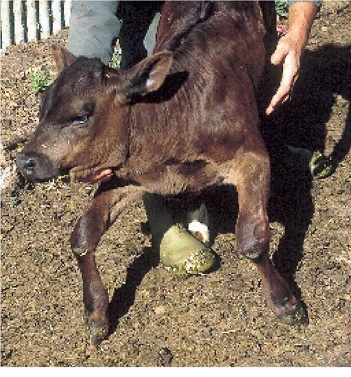
Plate 9.69. Ifthe legs are also flexed at the knee, the chances of recovery are much less.
More on the topic LAMENESS DUE TO LEG DISORDERS:
- Diaphragmatic disorders are usually congenital, though acquireddefects due to direct trauma or neurological injury (diaphragmatic palsy) are not uncommon.
- Leg Length Inequality
- OTHER CAUSES OF FOOT LAMENESS
- The term congenital myopathy is used to describe a group of heterogenous disorders usually presenting with infantile hypotonia due to genetic defects, causing primary myopathies with the absence of any structural abnormality of the central nervous system or peripheral nerves.
- FOOT CONDITIONS CAUSING LAMENESS
- Nutritional anemia is defined as the anemia due to deficiency of one or more micronutrients required for normal erythropoiesis and mainly includes: (a) iron deficiency anemia, and (b) megaloblastic anemia, due to vitamin B12 or folic acid deficiency.
- CHAPTER 9 LAMENESS AND FOOT TRIMMING
- Lameness is not only a major economic problem, but it is also a major welfare issue - for both the cow and the herdsman!
- P Keerthi Kundana, Mona Gajre, Alpana Kondekar, Mukesh AgrawalNeurological disorders account for ~15-20% of hospitalizations, which may be divided into three major categories: (a) central nervous system disorders, involving brain and spinal cord, (b) neuromuscular disorders involving peripheral nerves and muscles, and rare disorders of autonomic nervous system.
- Human TB Due to M. bovis