OTHER CAUSES OF FOOT LAMENESS
I have dealt extensively with sole ulcers and white line disorders because they are two of the most important causes of lameness and because their control is so complex. Other causes of foot lameness are:
Hoof disorders
foreign body penetration
slurry heel
haematoma in the heel vertical fissures (sandcracks) hardship lines and coriosis horizontal fissures broken toe
Skin disorders
interdigital necrobacillosis (foul or footrot)
digital dermatitis (hairy warts) and interdigital dermatitis interdigital skin hyperplasia (corns, growths or tylomas)
mud fever
Bone and joint disorders pedal bone fracture pedal bone tip necrosis pedal arthritis
Foreign Body Penetration of the Sole
Typical foreign bodies are stones (especially sharp flints), nails (particularly those with flat heads), fragments of wood, glass or tin, and occasionally even the sharp root of a cast tooth will penetrate the sole.
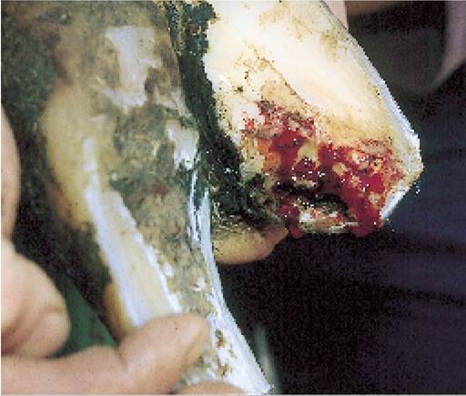
Treatment is very similar to that for white line disease (page 299), namely remove the foreign body and then all under-run horn. In Plate 9.39 a nail has been removed but obviously there is still under-run horn towards the heel at A. This must be pared away to allow new horn formation on the underlying corium.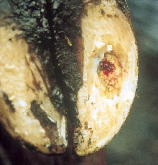
A
Plate 9.39. Puncture of the sole by a foreign body. The red area of corium is the initial point of penetration. There is still more under-run sole to be removed at A.
Slurry Heel
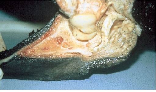
Plate 9.40. Advanced slurry heel removes support from the pedal bone which then pinches the underlying corium. An external view of slurry heel is shown in Plate 9.47.
The smooth, soft and pliable horn of a normal heel can be seen in Plates 9.23 and 9.24.
In feet which have been exposed to slurry over a long period of time, the heel horn often becomes black and pitted and in more extreme cases totally eroded, as in the foot with digital dermatitis (Plate 9.47). Although perhaps not looking too serious from the outside, slurry heel causes important internal changes. Removal of weightbearing at the heel allows the foot to rotate backwards, thereby predisposing to sole ulcers, as explained in Figure 9.15. Plate 9.40 shows an advanced case in which the flexor tuberosity at the rear of the pedal bone no longer has adequate support and as a result is penetrating the horn of the sole. The corium at this point will be pinched every time the cow walks. Slurry heel is controlled by keeping the feet clean and dry, using lime in cubicles, frequent scraping of cubicle passages and footbaths (page 318).Haematoma in the Heel
A haematoma (blood blister) in the heel is almost certainly a result of trauma. Most cases occur in cows walking to and from grazing. Uncomplicated cases produce only a slight swelling of the heel bulb and mild lameness, and can be treated by incising the heel and draining the blood (as would be done for similar damage to a human finger-nail). In some cases the haematoma develops into an abscess or may even lead to necrosis and a total slough of heel tissue. More extensive drainage and use of a shoe on the sound claw are then required.
Vertical Fissures (Sandcracks)
Vertical fissures occur as a result of damage to a small area of the periople and underlying coronary band. Horn formation is then disrupted at that point, although the adjacent horn continues to grow. This leaves a gap (the vertical fissure) running down the hoof wall from the point of disrupted production (Plate 9.41). In North America vertical fissures are commonly seen in both grazing beef cattle and older dairy cows kept in sand lots, where the combination of age, sand, wind and dry weather removes the protective periople. Vertical fissures can also occur as a result of a digital dermatitis infection on the coronary band (Plate 9.50).
Supplementation of the diet with biotin (10 mg per cow per day) may help to prevent fissures.For treatment, pare out the fissure using the curved tip of the hoof knife. A small abscess may be found under the wall, as in Plate 9.42. If the fissure is large and runs full length, apply a block as in Plate 9.50.
Hardship Lines
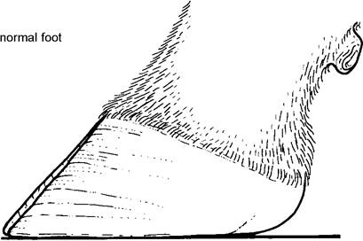
Any disruption in horn formation may leave a groove, sometimes referred to as a hardship groove, encircling the hoof wall. These are the result of coriosis/laminitis. Inflammation of the laminae leads to massive pressure under the hoof wall, causing the wall to push forward and the toe to lift, as in Figure 9.21. The eventual effect is a concave front wall with numerous hardship lines. In Plate 9.4 note the obvious bands of hardship lines running parallel to and just down from the coronary band.
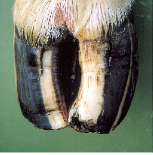
Plate 9.41. Avertical fissure is a split running down the front wall of the hoof.
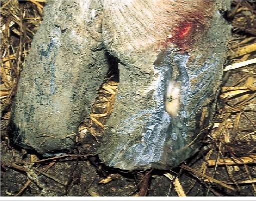
Plate 9.42. A small abscess in the laminae of the corium beneath a vertical fissure made this cow extremely lame.
Horizontal Fissures
If an animal is severely ill, for example with mastitis, metritis or any toxic condition, there may be a total cessation of horn formation for a while. When horn production starts again, instead of there being a hardship groove, there may be a horizontal fissure completely encircling the hoof wall. Initially this may cause no problem, but as the defect grows down towards the toe it loses its support and attachment from the heel. The protruding ‘thimble’ of horn is then able to move on the underlying corium, causing pinching, pain and lameness. A typical example is shown in Plate 9.43. This cow had been badly affected by foot- and-mouth disease some three months previously and as a result had a horizontal fissure on the claws of all four feet.
The date of the illness can be calculated by measuring the distance from the coronary band to the fissure (approximately 15 mm) and dividing this by the rate of horn growth, namely 5 mm per month: 15 divided by 5 = 3 months.For treatment, remove the loose ‘thimble’ of horn. If the corium is extensively exposed, apply a block to the sound claw. However, beware: not all horizontal fissures lead to lameness. Some simply grow to the toe and are shed naturally. It is only necessary to trim the foot if the cow is lame. A cow with one long claw and one short, due to the horizontal fissure fragment having been shed from one claw only, is sometimes referred to as having a broken toe. This is shown in Plate 9.44.
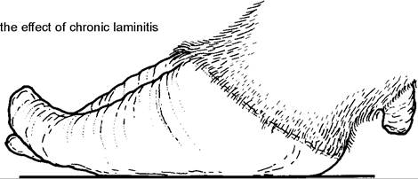
Figure 9.21. Laminitis distorts claw growth and may produce hardship lines, a concave front wall, an upward rotation of the toes and sinking of the heel.
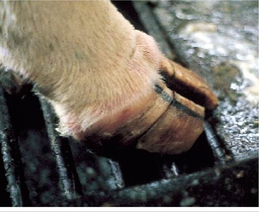
Plate 9.43. A horizontal fissure results from a total, but temporary, cessation of horn formation, usually caused by illness (in this case foot-and-mouth).
Interdigital Necrobacillosis (Foul, Lewer, Foot Rot)
This is a bacterial infection of the interdigital cleft, caused by two organisms:
• Bacteroides melaninogenicus initially penetrates the skin surface and allows the entry of the secondary organism, namely:
• Fusobacterium necrophorum invades and its necrotising toxins destroy the deeper tissues of the dermis which causes the lameness.
Disease may be seen in both young calves and adult animals. Initially there is swelling of the foot, which typically pushes the claws apart. Soon after, the skin between the claws splits (Plate 9.45) to reveal pus, necrotic debris and sometimes blood. Some say that there is a characteristic smell.
In untreated cases the infection may track up the tendon sheaths of the leg, or penetrate the pedal joint, both producing severe lameness.Treatment is simply antibiotic by injection, but the foot should always be checked to ensure that there is no stick or stone present penetrating the interdigital skin. For control, ensure that cattle are not exposed to sticks, stones or thorns which might
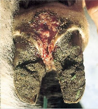
Plate 9.46. ‘Super foul’ is a colloquial term applied to an extremely virulent form of the disease which produces severe damage.
Plate 9.44. The thimble of loose horn beyond a horizontal fissure is sometimes referred to as a broken toe.
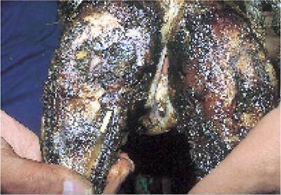
Plate 9.45. Interdigital necrobacillosis (foul, footrot, lewer) is recognised as a split in the skin between the claws.
damage the interdigital skin and make sure that areas around water and feed troughs are kept clean, as this is an area where infection can be transmitted from cow to cow. Footbaths (page 318) can also help.
In the UK a new and highly virulent form, colloquially termed ‘super foul’, sometimes occurs (Plate 9.46). The damage caused to the foot in as little as 24 hours is spectacular. No new strains of bacteria have been isolated, but most affected herds have a concurrent digital dermatitis infection which probably allows the entry of higher challenge doses of the ‘foul’ organisms. Prompt and prolonged antibiotic (for five to seven days or more) is needed for treatment. It has been suggested that strapping clindamycin or a similar antibiotic which is effective against anaerobic bacteria into the interdigital cleft may also help.
Digital Dermatitis (Hairy Warts)
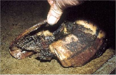
Digital dermatitis is another bacterial infection of the skin, but this time only the surface layer (the epidermis) is involved. It is caused by a spirochaete, probably a member of the Treponema family, but
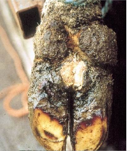
Plate 9.47.
Digital dermatitis is seen as a moist, painful, smelly area, grey or red, radiating out from the interdigital pouch. Slurry heel is also present. despite the worldwide incidence of the disease the organism has yet to be precisely identified.Early cases are typically seen as a moist, light greyish-brown area of exudate, with matted hairs, situated on the skin between the heel bulbs at the back of the foot (Plate 9.47). Cleaning the lesion reveals:
• a red, raw or necrotic area radiating out from the interdigital pouch. This pouch is at the rear of the interdigital cleft and often acts as a reservoir of infection
• a characteristic pungent, sulphur-like smell, thought to be caused by the Treponema bacteria decomposing the keratin within the skin
• intense pain, surprisingly so for what appears to be a relatively mild lesion
The above is a description of a typical acute lesion.
Chronic, longstanding cases produce the syndrome of hairy warts (Plate 9.48), common in North America but rarely seen in the UK. The filaments of these warts are in fact lengths of epidermis produced by the skin growing as fast as possible in an attempt to shed the organism from its surface. Because of their chronic nature, hairy warts are more difficult to treat than ‘standard’ digital dermatitis.
Although digital dermitis typically radiates from its reservoir in the interdigital pouch (as in
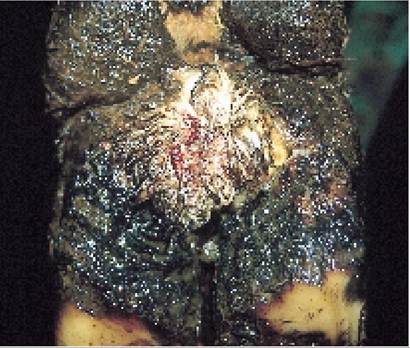
Plate 9.48. Hairy warts is a chronic form of digital dermatitis. The filaments of the warts are fronds of skin.
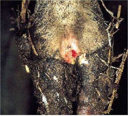
Plate 9.49. Digital dermatitis can also occur at other sites on the foot, including the coronary band.
Plate 9.47), lesions may be seen at many other places on the foot, for example:
• across one heel and spreading up towards the accessory digit
• under-running the sole from the heel
• at the front of the foot (Plate 9.49). Disease is particularly dangerous at this site. Involvement of the coronary band tissue can produce a total vertical fissure (Plate 9.50) leading to protracted lameness
• between the claws. Some books say that this is a separate condition, known as interdigital dermatitis, but its appearance, smell and response to topical antibiotics make it highly probable that it is digital dermatitis in a different site. Plate 9.51 shows digital dermatitis on the surface of a corn
• occasionally as a secondary infection to sole ulcers
Treatment of digital dermatitis consists of cleaning the lesion and applying topical antibiotics. Lincospectin and oxytetracycline are most commonly used and repeated topical applications are beneficial. With the more chronic form of hairy warts a dressing impregnated with antibiotics may have to be strapped in position for several days. For anterior lesions involving the coronary band, both injectable and topical treatments should be used.
Digital dermatitis is a disease associated with larger groups of cattle in conditions of close confinement, high stocking density, damp conditions and poor foot hygiene. Control measures are therefore based on the following:
• Scrape cubicle passages and feed areas at least twice a day, making sure that all stale slurry is removed from places like water troughs and feed areas.
• Keep feet as dry as possible. The use of lime in cubicle beds will help, as will the luxury straw levels depicted in Plates 9.35 and 9.36. Part of the straw will be pulled out into the cubicle passage, further reducing the exposure of feet to slurry.
• Give the whole herd antibiotic treatment. This can be applied either as a jet onto the heel of each cow, for example via a garden water sprayer, or by walking the cows through an antibiotic footbath (page 318). Usually once a month is sufficient.
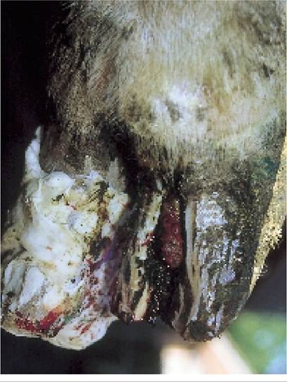
Plate 9.50. Avertical fissure may result from digital dermatitis affecting the coronary band.
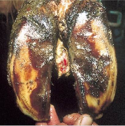
Plate 9.51. Interdigital skin hyperplasia (corns, tylomas) is caused by some factor irritating the skin between the claws. In this instance digital dermatitis is present on the surface.
Regular footbaths are even more important for the chronic form of hairy warts. Although they may not always cure affected cases, frequent baths will help to prevent the establishment of new and chronic cases. Over the course of two or three years of treatment of clinical cases and prevention of new cases by the use of a two- or four-weekly footbath, the syndrome should eventually come under control. A degree of immunity to digital dermatitis must develop, because in chronically infected herds the disease is most commonly seen in recently introduced heifers or in purchased cows two to six weeks after entry to a herd.
Interdigital Skin Hyperplasia (Corns, Tylomas, Fibromas, Growths)
As its technical name suggests, this is an overgrowth of normal skin originating from a natural fold in the interdigital cleft (between the claws). Atypical example is shown in Plate 9.51. In some cows, especially the heavier breeds, it can be hereditary. In others it is caused by chronic skin irritation, for example, from low-grade foul, digital dermatitis or simply impaction with dirt. Lameness is caused by the skin growth being squeezed by the claws during walking, or by the secondary infection of the growth with digital dermatitis or foul.
Mild cases can be treated by simply removing horn from the inner edges of the sole, adjacent to the sole ulcer site. This eliminates the pinching effect and the ‘growth’ then slowly disappears on its own. Larger lesions require surgical amputation.
Mud Fever
Mud fever occurs following exposure to cold, wet and muddy conditions. The lower leg becomes slightly swollen, with dry, hard and flaking skin. There may be hair loss (Plate 9.52) and even bleeding if the skin cracks. For treatment, thoroughly wash the legs. Dry and then apply a greasy antiseptic ointment or a teat spray which contains a high level of emollient. As the organism Dermatophilus may be involved, three days of injectable antibiotic (penicillin) may also help.
Fracture of the Pedal Bone
Bulling activity, with the mounting cow falling back heavily onto hard or rough concrete, is the most common cause of a fractured pedal bone (viz the bone inside the hoof - Figure 9.1). Bones weakened by age, fluorine poisoning or a foreign body penetrating the sole of the hoof may be more at risk of a fracture. Typically it is the inner claw of the front foot which is involved, and by adopting a cross-legged stance, as in Plate 9.53, the cow transfers her weight onto the sound lateral claw. However, the stance alone is not sufficient to diagnose fracture of the pedal bone. Cows with ulcers in both inner claws will adopt the same position. Most animals heal well if a block is applied to the sound claw.
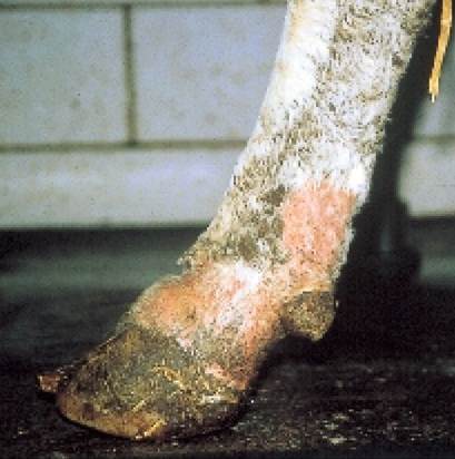
Plate 9.52. Mud fever occurs following prolonged exposure of the skin to wet and muddy conditions.
Plate 9.53. A cross-legged stance is said to be typical of a fractured pedal bone, but it can also occur if there are ulcers on both medial claws.
Pedal Bone Tip Necrosis
In a few cows, what initially appears to be a standard white line abscess at the toe sometimes fails to heal, even though it may have been treated thoroughly. At the second examination there will probably be a characteristic foul odour and even with further extensive removal of under-run tissues the toe fails to heal. A typical example is shown in Plate 9.54. Note how short the affected claw has become, compared to the normal claw on the left. In such cases the front tip of the pedal bone has become infected (technically known as osteomyelitis) and unless the damaged and infected bone is all removed, the claw will never heal.
Each time you pare the toe, it seems to go back even further. This is because more of the tip of the pedal bone has been eroded. The only treatment for such cases is either total removal of the digit or, using a wire, sawing off the tip of the toe to remove all the necrotic bone.
Pedal Arthritis
Inflammation of the corium produces changes in the horn leading to disorders such as sole ulcers and white line disease. The corium also feeds the pedal bone and so an inflamed or infected corium, perhaps caused by a severe longstanding ulcer or white line infection, can also produce internal changes on the bone. The example shown in Plate 9.55 is quite mild, but imagine how the protruding spicules of bone (A) will impact on the joint surface to make walking uncomfortable. Cows which develop chronically enlarged claws following a longstanding sole ulcer will have much more severe changes than this. There is no treatment.
An infected pedal joint (purulent arthritis) is an even more serious condition. It is usually the result of a
Plate 9.54. Necrosis of the pedal bone. Note how short the affected claw is compared to the normal one.
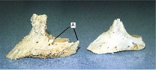
Plate 9.55. Mild pedal arthritis. Spicules of bone (A) protruding from the claw on the left will make weightbearing uncomfortable. A normal pedal bone is on the right.
severe or neglected case of foul, white line disease or sole ulcer. The foot becomes grossly swollen and infection may start to track up the tendon sheaths of the leg. The cow will be intensely lame, probably not using the leg at all. A typical example is shown in Plate 9.56. Radical treatment such as digit amputation and prolonged, aggressive antibiotic therapy may sometimes be effective. Many cases have to be culled.
More on the topic OTHER CAUSES OF FOOT LAMENESS:
- FOOT CONDITIONS CAUSING LAMENESS
- CHAPTER 9 LAMENESS AND FOOT TRIMMING
- LAMENESS DUE TO LEG DISORDERS
- FOOT TRIMMING
- Lameness is not only a major economic problem, but it is also a major welfare issue - for both the cow and the herdsman!
- FOOT-AND-MOUTH DISEASE
- THE STRUCTURE OF THE FOOT
- Immersion Injury (Trench Foot)
- An Infeasible Assumption and Limited Information Hinder Veterinarian Workforce Planning Efforts for a Catastrophic Outbreak of Foot-and-Mouth Disease
- It was with “great wonderment” that Peter I learned of “the deed of the new Judas, Mazepa, who, after twenty-one years of loyalty to me and with one foot already in the grave, has turned traitor and betrayer of his own people.
- American religious history began some 30,000 to 50,000 years ago when, according to archaeologists, the first human beings set foot on the North American continent.
- Feet
- CAUSES OF DISEASE