SOLE ULCERS AND WHITE LINE DISEASE
As the causes of sole ulcers and white line disease are very similar, these two conditions will be dealt with in the same section. Aknowledge of the pathogenesis (the internal changes) leading to sole ulcers and white line disease will help us to appreciate the structure and function of the foot and is described in the following section.
This knowledge also considerably improves our understanding of the control measures necessary.Coriosis (Laminitis)
On page 281 we saw that the horn of the sole was produced by the corium of the sole. Therefore, if the corium becomes damaged or inflamed, horn formation is likely to be changed in some way. Although, as mentioned previously, we often refer to ‘laminitis’ as meaning inflammation within the foot, in many instances it is either the whole corium which is inflamed, or just the corium of the sole. The term ‘coriosis’ is therefore more likely to be correct.
Inflammation and damage to the corium can be the result of a range of things, for example:
• trauma
• infection
• nutrition and metabolic disorders
• toxins
However, the overall result will be the same, namely altered horn production.
Sole haemorrhage and bruising
Inflammation of the corium leads to increased blood flow. This produces congestion in some areas, with pooling of blood and poor oxygenation leading to tissue damage and poor horn formation in others. The whole process results in the corium becoming much more fragile. In the early stages serum (fluid)
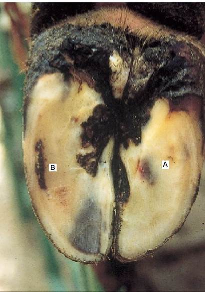
Plate 9.20a. Coriosis: blood released into the horn is seen on the surface of the sole one to two months later. On this foot there is haemorrhage at the ulcer site (A) and the white line (B).
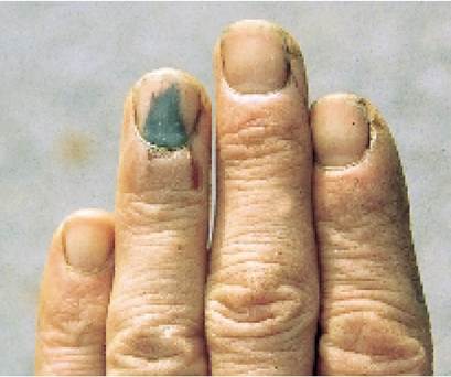
Plate 9.20b.
Coriosis: a bruise on a finger (here caused by the author foolishly putting his hand into a cow's mouth without a gag!) grows down along the nail in an identical manner to sole bruising. leaking from the blood vessels within the inflamed corium is mixed with the horn of the sole as it is being formed. In more advanced cases, rupture of the capillaries produces a mixture of horn and blood. The sole is 5-10 mm thick, so with horn growing at 5 mm each month, it will take one or two months for this deformed and damaged horn to reach the surface. A typical example is shown in Plate 9.20A. There is blood mixed with the horn at both the sole ulcer site A and in the white line B. On the sole adjacent to B especially, the horn has a yellow appearance, due to leakage of serum into the horn. Note that the wall of the hoof adjacent to B is still a good, white colour. This horn is considerably older, having been produced at the coronary band several months previously, and so far has not been affected.Haemorrhage on the sole, seen in Plate 9.20A, is often referred to as bruising. This may be a correct term, although it should always be remembered that the ‘bruise’ was formed by an insult one or two months previously, when the horn now on the surface of the sole was being produced. As such, bruising of the sole cannot be implicated as a recent cause of lameness.
The effect of mixing serum or blood with the horn can be likened to mixing sawdust with concrete - it weakens it considerably. This is particularly the case with the white line, which is an inherently weak structure, and at the sole ulcer site where there may be almost ‘neat sawdust’ because so much haemorrhage is present. The whole process is very similar to the changes which occur when your finger-nail is bruised (Plate 9.20B): the blood spot often starts at the corium of the skin-nail junction and then slowly grows to the tip of your nail over the next few months.
Changes associated with the pedal bone
In Plate 9.3 we saw that the inner border of the pedal bone is arched in shape.
The pedal bone is suspended within the hoof by the laminae, with a much stronger attachment to the outer wall than to the inner one. When weight is transmitted down the leg the bone rotates slightly inwards, putting increased weight on the flexor tuberosity, which is the rear projection of the pedal bone (F in Figure 9.15 and Plate 9.3). Increased weightbearing at this point puts extra pressure on the corium and if it is already in a fragile state, then it is even more likely to become damaged. Pinching of the corium between the pedal bone above and the horn of thesole beneath can lead to bruising, as shown in Figure 9.15 and Plate 9.20A. This bruising will appear on the surface of the sole one or two months later and may be seen as:
• yellow discolouration - if only serum was released
• haemorrhage - if the blood vessels ruptured
• a sole ulcer - if the damage to the corium was so severe that horn formation was totally disrupted
If there is a generalised inflammation of the corium, the suspension of the pedal bone within the foot is disrupted, allowing the bone to sink within the foot, as shown in Figure 9.16. This further complicates the situation by producing:
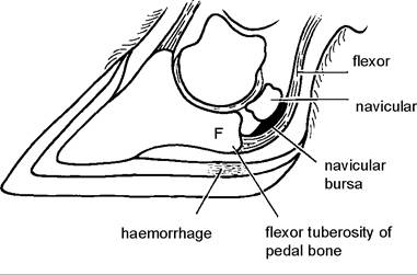
Figure 9.15. Sole ulcer formation. Pinching of the corium between the flexor tuberosity (F) of the pedal bone and the horn of the sole leads to release of blood into the horn.
• both sole and toe ulcers
• permanent poor horn formation
• expansion and weakening of the white line
• swelling around the coronary band
As the pedal bone sinks within the hoof it displaces the corium out to the side, as shown in Figure 9.16. This produces a very wide, weak white line and a very large and flattened sole. Sometimes the corium is displaced so far to the side that the wall curves outwards. I find such feet particularly difficult to trim. On the one hand you want to bring the claw back to its correct shape, but in so doing it may be necessary to remove all the weightbearing wall.
The sinking pedal bone may also displace part of the corium upwards. The upward displacement is seen as a thickened ring of swollen tissue, running around the hoof just above the coronary band, as in Plates 9.18 and 9.19.As the pedal bone sinks onto the corium, sometimes the front part of the bone sinks before the rear, producing haemorrhage at the toe. This is clearly seen on the left claw in Plate 9.21 and is often referred to as a toe ulcer (A). Note how there is also haemorrhage (H) in the white line towards the heel on both claws, extensive yellow discolouration of the sole due to serum infiltration and an early sole ulcer (S).
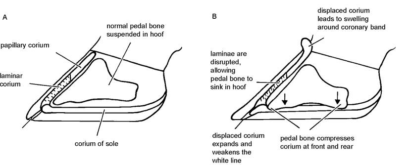
Figure 9.16. In the normal claw (left) the pedal bone is suspended within the hoof by the laminae. Coriosis/laminitis may disrupt this suspension (right), allowing the pedal bone to drop onto the sole, thereby further compressing the corium (courtesy Dr. P. Ossent).
However, it is more usual for the rear edge of the pedal bone to sink within the hoof, pinching the corium under the flexor tuberosity and producing the classic sole ulcer.
Continual compression of the corium of the sole can lead to generalised poor horn formation, sometimes seen in older cows where a sole ulcer fails to heal totally. A layer of very poor-quality horn may form over the ulcer site, and the tip of the flexor tuberosity of the pedal bone can sometimes be palpated as a hard lump just below. Once the pedal bone has sunk within the hoof, it is unlikely ever to regain its original position. This is why it is so important to prevent the development of sole ulcers in heifers.
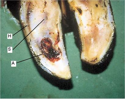
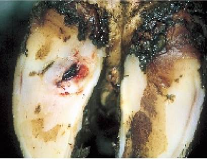
Plate 9.22. Sole ulcer, early stage. Note the discharge of fresh blood and serum at the ulcer site.
Plate 9.23. Sole ulcer, later stage. The ulcer site has turned black and necrotic.
Plate 9.21. Atoe ulcer is produced when the front tip of the pedal pinches the corium. (A- toe ulcer, S - sole ulcer, H - white line haemorrhage.)
Sole Ulcers
A sole ulcer is formed when damage to the corium is so severe that there is total disruption of horn formation. If the foot is pared in the early stages, before the ulcer appears, removal of the surface layers of sole horn may expose a soft area of moist, damaged horn, and yellow/brown fluid will run out (Plate 9.22). This is serum. At this stage there is no infection present, just physical damage. When the damaged horn has worked its way to the surface, infection enters from the environment and the area turns black and necrotic, as in Plate 9.23.
A sole ulcer is a physical condition, caused by trauma, and treatment must be aimed at reducing this trauma. The main steps for treatment are:
• Dish the sole ulcer site so that weightbearing is minimised.
• Remove any under-run horn around the ulcer, to eliminate pockets of necrotic horn and infection, thus allowing the formation of new horn.
• Remove any protruding granulation tissue (shown in Plate 9.24).
• Reduce the size of the affected claw as much as possible, so that weightbearing on the sound claw is maximised.
The use of blocks is an excellent treatment and is described on page 319.
On occasions, severe or neglected ulcers (or white line abscesses) may allow infection to
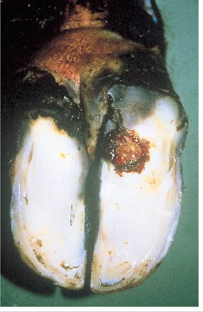
Plate 9.24. Sole ulcer, with granulation tissue protruding from the damaged corium.
penetrate into some of the deeper structures within the foot. Examination of Figure 9.15 shows that a sole ulcer lies immediately beneath the point of attachment of the flexor tendon to the pedal bone.
Small fragments of white, fibrous tissue can sometimes be seen protruding from deep ulcers. An example is shown in Plate 9.58. These are pieces of flexor tendon. If infection is allowed to progress, there may be total rupture of the tendon, to leave the toe permanently rotated, as in Plate 9.25. Penetration into the deeper structures, such as the navicular bursa or even the pedal joint itself, produces a very severe lameness, with a swollen claw and purulent discharge from the ulcer site, as in Plate 9.26.Radical treatment is now needed, perhaps using a block (as in Plates 9.26 and 9.58), flushing the abscess, or possibly total amputation of the digit. A block, a very large drainage hole and a long course
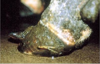
Plate 9.25. Sole ulcers which damage the attachment of the flexor tendon to the pedal bone result in a permanent upward rotation of the toe.
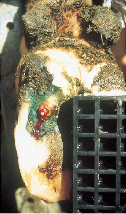
Plate 9.26. Aswollen claw and pus discharging from the ulcer site are a clear indication that infection has penetrated deeper structures within the foot.
The internal changes within the foot which lead to sole ulcers and white line diseases are:
• pinching of the corium between the pedal bone and the horn of the sole
• increased fragility of the corium
• disruption of the pedal bone suspension, allowing it to sink into the hoof
• lateral displacement of the corium of the sole into the white line area and dorsally to the coronary band
Plate 9.27. White line separation, with a penetrating stone. The stone may be shed by the normal growth of the hoof, or may penetrate deeper until it reaches the corium.
of injectable antibiotics at a high level (for example 7-10 days) will often be successful. Although digit amputation can work well, additional time and effort are needed for regular dressing of the foot, and considerable strain is imposed on the remaining claw.
Heel and Toe Ulcers
Although sole ulcers are by far the most common, there can be areas of haemorrhage or even total perforation at other areas of the sole. Toe ulcers are thought to occur when the pedal bone sinks within the hoof ‘bows first’, that is the front of the pedal bone drops before the flexor tuberosity at the rear (Plate 9.2l). Heel ulcers (sometimes referred to as ‘necrotic heel tracts’) are seen as small dark red/black marks in the central sole area towards the heel. Some simply track down to the corium and fade to nothing. Others lead to under-running of horn at the sole-heel junction and can produce a marked lameness. The cause of these heel ulcers is not known, although one theory is that they are produced by a pinching of the corium under the rear edge of the pedal bone.
White Line Diseases
Weakening of the white line, brought about by the inflammation associated with laminitis/coriosis, can result in a range of white line disorders. The most common of these are:
• sterile abscessation
• white line separation
• white line penetration and abscess
Sometimes the internal inflammation within the foot is so severe that pockets of necrotic tissue are formed. These can produce a sterile internal abscess and as there may be no obvious tracks running from the outside, they may be quite difficult to locate and treat. In severe forms of coriosis the whole sole becomes separated by an accumulation of inflammatory fluid. When foot trimming you may have seen a total layer of sole separated from the new sole underneath. This is known as a false sole.
More commonly the weakened white line starts to open up, a process known as white line separation. This occurs especially if the cows are walking over rough or stony ground, or when they
make sudden turning movements, as when escaping from an aggressive cow. Small stones may then become impacted, as in Plate 9.27, and with continued walking these may eventually penetrate the corium.
The most common point for white line separation and penetration is on the outer wall, near to the heel, as in Plate 9.27 or point 3 in Figure 9.13B. During locomotion this is where there are the greatest sheer forces between the rigid hoof wall, the suspended pedal bone and the movements of the flexible heel. Once the corium has been penetrated, the invading foreign body (usually a stone or grit) introduces infection. The bacteria multiply to produce pus and the expanding pus then has to find the easiest way of escape.
For white line abscesses near to the heel, this escape route is usually through the soft horn of the heel, as in Plate 9.28. White line abscesses close to the toe do not have such an easy escape route and often infection tracks upwards through the laminae, to discharge at the coronary band, as in Plate 9.29. This produces a more severe lameness because, as shown in Figure 9.1, the pedal bone is tightly attached to the hoof towards the toe and there is therefore very little room for the pus to expand.
Whereas a sole ulcer results in damage to the underlying corium, the majority of uncomplicated white line lesions only produce separation of the horn from the underlying horn-forming corium. (Note: the word ‘lesion’ means any pathological change in a tissue. In this instance ‘lesion’ could be separation, haemorrhage, abscess etc.). White line lesions normally heal much more quickly, therefore, than sole ulcers. In Plate 9.28 you can see how pulling back the flap of sole horn with a hoof knife exposes a pink tissue. This is corium covered by epidermis and it will soon form another good layer of protective horn.
The treatment of a white line abscess is very similar to that for a sole ulcer, namely:
• Remove all under-run horn, even if this means removing the wall from the sole to the coronary band (as in Plate 9.30), or the whole of an under-run sole.
• Reduce the size of the affected claw, to minimise weightbearing, and leave the sound claw as large as possible.
Blocks and dressings are discussed on page 319.
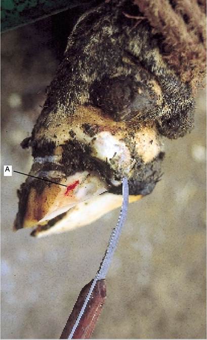
Plate 9.28. Many white line abscesses discharge at the heel. The original point of entry of infection is at A.
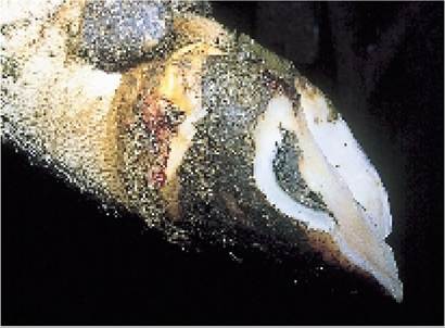
Plate 9.29. White line abscess discharging at the coronary band.
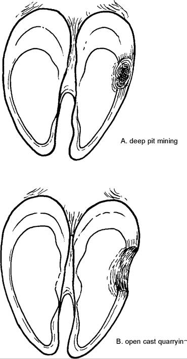
Figure 9.17. When searching for white line abscesses the technique should be one of ‘open cast quarrying' (B), not ‘deep pit mining' (A).
When using a hoof knife to drain infection from the white line, the approach should be one of ‘open cast quarrying’ rather than ‘deep pit mining’. This difference is shown in Figure 9.17. Digging a deep pit with the curved point of the hoof knife has two disadvantages, namely:
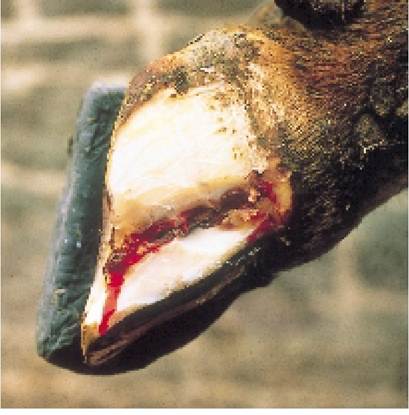
Plate 9.30. White line abscess treatment. All under-run horn must be removed.
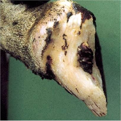
Plate 9.31. Protrusion of granulation tissue from the original white line abscess site probably means that there is further under-run horn. The coronary band is also swollen, suggesting infection of the deeper tissue.
• It leaves a pit which can easily become impacted with stones or dirt, thereby impeding drainage and predisposing to further white line impaction.
• By digging a pit you are much more likely to miss areas of under-run horn and pockets of infection.
If a small area of adjacent wall is removed, it is much easier to expose and drain the affected area. Complications can occur with white line abscesses, particularly those which track up the wall (e.g. Plate 9.30). Plate 9.31 is a typical example. Note the granulation tissue protruding from the original site of white line penetration and how the coronary band area is enlarged and inflamed.
Protruding granulation tissue is often an indication that there is adjacent under-run horn which needs to be removed. The swollen coronary band is probably caused by infection tracking into deeper structures such as the navicular bursa or tendon sheaths. A similar change is produced when the pedal bone ‘sinks’ onto the corium of the sole in acute coriosis (see Figure 9.16).
Causes and Control of Sole Ulcers and White Line Diseases
This is a huge subject, enough to fill a whole textbook on its own, and the reader must appreciate that only an outline can be given in this section. The internal changes within the foot leading to sole ulcers and white line disease were described on page 293. In the following the many environmental, managemental and nutritional factors which cause these changes is given. These could be listed in a variety of ways, but the system I have chosen, namely relating aetiology to pathogenesis, will, I hope, give a clearer understanding of how lameness is best controlled.
Earlier in the chapter I said that increased fragility of the corium predisposed to both sole ulcers and white line disease. So what causes increased fragility of the corium and what predisposes to damage? This will be covered under the following headings:
• calving
• excessive standing
• nutrition
• general management
Calving
There can be no doubt that calving (or maybe the start of lactation) is a major stressor on horn formation. We only have to look at the rings on a cow’s horns to see this. Plate 9.32 is a picture of Pinky, a thirteen-year-old cow from Figtree, Zimbabwe. She had only six calves in her thirteen years - not exactly a stressful life! - but note the six rings on her horns. There is one ring for each calving (although it is accepted that disease or periods of severe undernutrition can sometimes produce the same effect). Look at the bull’s horns shown in Plate 9.5: you will not see any rings present, irrespective of his age. There is something about calving which produces a disruption in horn formation and this occurs in both the horns and the feet. It also explains why the peak incidence of lameness occurs one or two months after calving: this is because it takes this length of time for the horn produced at calving to work its way down to the surface of the sole.
The natural decrease in rumination at the time of calving, leading to periods of rumen atony and potential acidosis, was discussed in Chapter 6, Figure 6.8. The importance of feeding long straw at this time to stimulate rumen contractions after
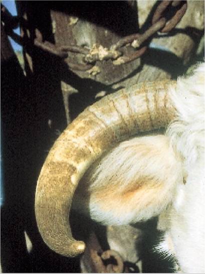
Plate 9.32. There are rings on a cow's horn, one for each calving. Disruption of horn formation also occurs in the claw. (Courtesy M. Conolly)
calving was also described. It is still not known why acidosis produces coriosis/laminitis, but bacterial endotoxins, leading to damage of the minute blood vessels (capillaries) within the foot, could be a factor.
It is not yet known why there is such a marked disruption in horn formation at calving. Suggested causes include:
• reduced rumination times
• an increase in acute phase proteins (for example, haptoglobulins)
• repartition of sulphur amino acids towards milk production
• cows (and especially heifers) spend longer standing immediately after calving
• the greater susceptibility of the cow to illness around the time of calving
Acute phase proteins are most commonly produced in response to disease. It is therefore interesting that the cow also experiences a marked rise in acute phase proteins at calving, especially as in the immediate postpartum period she is particularly susceptible to infection. Severe inflammation, caused by any disease, can disrupt horn formation and lead to horizontal fissures (page 312), so could calving simply be an extension of this process? We know that administration of cortisone to horses can induce laminitis/coriosis and also that the ‘signal’ to initiate the process of calving is cortisone produced by the developing foetus (Chapter 5). Could these processes be connected with lameness?
Calving also sees the start of lactation. As milk production rapidly rises there is an enormous increase in the demand for sulphur-containing amino acids, because many are essential for lactation. Recent work has shown that horn produced at the time of calving has a lower sulphur content. Sulphur is an important ingredient of keratin, the protein which leads to hardening of horn; therefore an inadequate supply of sulphur will lead to soft horn. This in itself would not be sufficient to cause the enormous damage seen in the feet of some heifers, but it could be a contributory factor.
Even if they calve outside in a field, for a few days after calving, cows and especially heifers will spend far more time standing and their lying times will be decreased. There is therefore more weight on the corium and a greater potential for bruising. It is not known whether the decreased lying times are due to nursing behaviour (attending to the calf), discomfort from the perineum (vulva or vagina), an enlarged udder or some other factor.
Diseases such as mastitis and metritis are certainly more common immediately after calving and as we know from hooves with horizontal fissures (page 312), acute illness affects horn formation. However, this will only involve individual animals.
Excessive standing
Anything which leads to a decrease in lying times, especially in the immediate post calving period when the corium is in its most fragile state, will increase the incidence of sole ulcers and white line disease. Heifers are likely to be the most greatly affected, and the worst case scenario of heifers entering a dairy herd which was described in the section on stress in Chapter 8 (page 268) should be read in conjunction with the following.
Increased lying times can be achieved by:
• maximising cubicle comfort, or using straw yards for the first few weeks after calving
• encouraging animals to enter cubicle houses
• training heifers to use cubicles during rearing
• minimising the time animals spend waiting to be milked and fed
• providing ample loafing and exercise areas
• avoiding overcrowding
One experiment deliberately housed heifers in an overstocked cubicle building (17 cubicles for 35 heifers) immediately after calving. Although the average lying time of the heifers was ten hours, some animals lay down for as little as five hours each day. This group showed the highest incidence of lameness, and quite severe haemorrhage persisted in the sole horn for up to four months after calving. In most dairy systems the heifers are forced to spend longer on their feet after calving. They will be waiting to be milked, they spend longer standing and feeding because they are often last to feed, and they need to eat more as lactation proceeds. They have recently been mixed with the main herd and are now having to compete with older cows. Fear may restrict their entry into a cubicle shed, especially if they are of low social dominance and have had no previous cubicle training.
Excessive standing may be bad for the immediate post-partum cow, but standing still is even worse. If the animal does not move around enough, the pumping mechanisms of the heel and digital cushion will be impaired, the blood will become ‘stale’, due to a lack of nutrients (particularly oxygen), and tissue damage, with poor horn formation, will result. It is essential that there are adequate loafing areas to allow the cows to walk around freely. Overcrowding should be avoided, even in collecting yards. Animals which are packed tightly together have little option but to stand still. Adequate loafing areas also help to improve fertility.
In summary, the incidence of sole ulcers and white line disease will be markedly reduced if animals are encouraged to maximise lying times in the immediate post calving period, for example, for the first two to six weeks.
Post calving comfort Of all the above factors, cubicle comfort is probably the most important. Cubicles may make the management of cows easy, but they are not always ideal in terms of cow comfort and lameness. For example, in a survey of dairy herds carried out by Edinburgh University, the incidence of lameness in cows housed in straw yards was only 5%, compared with 25% for cubicles. This must point to cubicles being less than ideal, especially for the immediately post calving cow. In fact a small but increasing proportion of farms are now housing their freshly calved cows in straw yards for the first two to six weeks after calving and then transferring them to cubicles. Experience from such systems suggests that in heifers especially, a post calving period of straw yard housing leads to:
• increased yields
• a decreased incidence of lameness
• improved cubicle acceptance when the heifers are eventually transferred from the straw yard to the cubicles
The third factor is perhaps the most surprising. One might have expected that cows and heifers which had got used to a straw yard would be very difficult to retrain to use cubicles. The fact that the reverse is true probably tells us that calving is a much more stressful experience than we think and that it is only when the cows have fully recovered that they are able to withstand the rigours of the cubicle system.
Cubicle design Cubicle comfort is obviously all-important. Ideally, cubicles should be long enough and wide enough (1.15 m wide and 2.4 m long) to accommodate the larger Holstein cows and with sufficient space at the front to allow the cow to lunge forwards 1-1.5 m as she stands up. If there are two facing rows of cubicles, a length of 2.2 m is adequate.
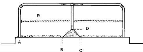
Figure 9.18. Cubicle design is important for comfort. They should be 1.15 m wide. Acentral concrete triangle BCD helps to position the cow correctly. A flexible rope R improves acceptance. (Courtesy John Hughes)
A good design is shown in Figure 9.18; there is a wide range of other designs which may be equally comfortable. This has a 100 mm fall from front to rear, a step of not more
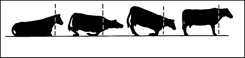
Figure 9.19. When rising, a cow lunges forward 1-1.5 m and puts enormous weight on her knees.
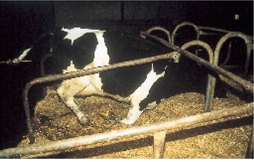
Plate 9.33. As a cow lunges forward to stand, she places enormous weight on her knees. The front of the cubicle therefore needs to be very well bedded.
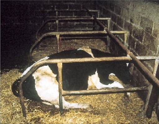
Plate 9.34. These cubicle beds were constructed of stone and only a thin layer of straw. In trying to get comfortable, this cow shuffled so far forward to the front wall that she was unable to stand up.
than 130 mm down into the dunging channel and an interesting concrete pyramid at the front. This pyramid prevents the cow from shuffling too far forwards, but at the same time provides ample space for lunging as she stands up. When rising she may place one foot on the slope of the concrete, to push herself up, but when fully standing she will have to keep her feet behind B and will then defaecate in the dunging channel. A neck rail is often not necessary and this may further increase cubicle comfort. The flexible rope division (R) eliminates pelvic damage which could occur with a solid central rail and it also avoids compression of the rumen.
If sitting in the cubicle means that the cow’s rumen is excessively compressed, or if there is insufficient space for her to extend her neck whilst regurgitating the cud, the cow is more likely to stand up to chew the cud, rather than do it lying down. Again, this will increase trauma to the feet. Cubicles with a high step (greater than 130 mm) from the dunging passage, with low bottom rails and with limited lunging space, have all been associated with increased lameness.
When attempting to stand, the cow lunges forwards 1-1.5 m and lifts herself first on to her hind feet, then up on to her front feet (Figure 9.19 and Plate 9.33). When she is lying down or half standing, therefore,
much of her weight is taken on her knees. If the floor surface is hard under her knees and particularly if it is also rough, cubicle acceptance will be low. The worst possible cubicle floor is a stone base, poorly compacted and with insufficient straw. This was provided for the cow in Plate 9.34. In an attempt to get comfortable she kept shuffling forwards - until she was so far forwards and so close to the wall that she was unable to stand. In the struggling, her back legs came forwards under her and by the time she was found in the morning such severe muscle damage had developed that she never stood up again. A cow lost, simply because the cubicle was uncomfortable. (There was also a very high incidence of lameness in this herd.) If you are finding a proportion of your cows stuck too far forward in the cubicles, reexamine cubicle comfort.
Most cubicle bases are made of concrete. This is fine provided the cubicle is deeply bedded, although
it is often difficult to get the straw to stay in. If this is the case, first put a 150 mm layer of rotted muck (for example, from the calf shed) onto the bare concrete at the front of the cubicle and then put clean straw on top of it. ‘Composted’ bedding from a straw yard does not have a particularly high E. coli level and its use does not predispose to mastitis, provided that plenty of clean straw is added to the top. It dries quickly and forms a good bed which adheres to the base of the cubicle. A variety of mats are available and these are certainly much better than concrete alone. However, some bedding should be used, even with mats; otherwise hock sores will develop (Plate 9.64). A disadvantage of mats is that it is difficult to get large amounts of straw bedding to adhere to them, although the cows enjoy standing on them.
The best cubicles are comfortable cubicles and if you can make them like mini-straw yards, so much the better. While design and dimensions may be important, I am convinced that comfort is of even greater significance. The cubicles illustrated in Plate 9.35 are an example. Although these cubicles measured only 1.07 m by 2.05 m and housed large FriesianZHolstein cross cows, they were regularly bedded as shown, with straw 380 mm deep, up to the bottom rail! Needless to say, they were extremely comfortable and as a result, the incidence of lameness was minimal. Another design of high comfort cubicles, deeply bedded and having a highly flexible division, is shown in Plate 9.36.
Plate 9.35. Luxury cubicle bedding. Although the cubicles are not ideal dimensions, the use of large quantities of straw made them very comfortable. (Courtesy M. Boynton.)
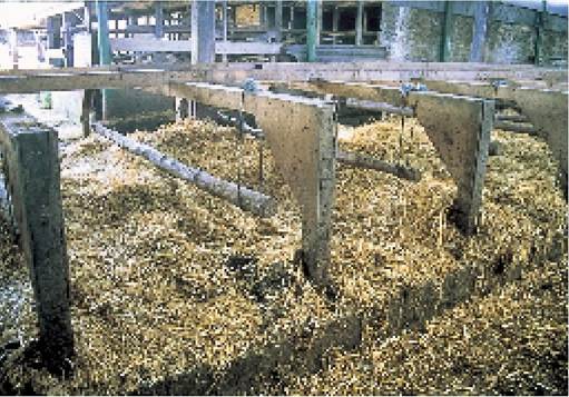
Plate 9.36. Luxury cubicle bedding with flexible dimensions for even greater comfort. (Courtesy R. Troughton.)
Nutrition
Although it is not easy to prove experimentally, diets which lead to acidosis undoubtedly predispose to coriosis/laminitis and subsequent lameness. The type of diet likely to cause acidosis and the prevention of dietary problems in general was discussed in Chapter 6.
Concentrate intakes should be built up slowly after calving, to reach a peak no earlier than two weeks post calving for average yielding cows and probably three weeks for higher yielding animals, which peak later. If there is a sudden change in diet to high concentrate feeding at calving, then problems may occur. For example, when the heifer whose foot is depicted in Plate 9.21 calved, her diet was immediately changed from an all forage (grazing) pre calving ration to a high fat, low forage out of parlour mix, together with 7 kg concentrate in the parlour. The resulting coriosis/laminitis produced severe lameness with both toe and sole ulcers and white line haemorrhage. Ideally no more than 4.5 kg of feed should be given in the parlour. The inclusion of 1-3 kg of long-chop straw, well mixed with the ration, helps enormously, in that it stimulates rumination, thereby promoting a good flow of saliva and decreasing acidosis. There is evidence that maintaining an ideal dietary cation-anion balance (DCAB, Chapter 6) may also help.
The most common dietary faults associated with lameness are:
• a sudden increase in concentrates after calving
• too much concentrate fed in a single feed in the parlour
• insufficient long fibre
• high starch and high oil
It is the composition of the ration and not its overall energy content which seems to affect the incidence of lameness. Table 9.1 shows two groups of cows, one of which (A) was fed a high fibre diet and the other (B) a low fibre and high concentrate ration. Both rations had the same overall crude protein (CP) content and both achieved the same energy (ME) intake, although the high fibre group clearly needed a higher dry matter intake to do so. The high incidence of coriosis/laminitis and sole ulcers in the low fibre group is striking. Despite regular foot trimming, group B also had a higher incidence of solar overgrowths (as in Plate 9.7). Although high protein diets have occasionally been suggested as a cause of coriosis, most people consider protein to be of less importance than other factors.
High intakes of poorly fer- Table 9.1. Two groups of cows having the same total daily protein mented grass silage have been energy intake, but Group Awas fed a high fibre diet and Group B a implicated, although this could low fibre diet.
| be due to toxic amines rather than high protein. | No. showing | ||||
| Even feeding during rearing | clinical | No. | |||
| influences the incidence of sole | ME | CP | coriosis/ | showing | |
| haemorrhage, with heifers fed high levels of concentrate being the worst affected. As discussed | Group A: | (MJ/kg) | (g/kg) | laminitis | sole ulcers |
| in Chapter 4, high fibre diets are now recommended for rearing | 26 cows fed a | ||||
| dairy heifers. | high fibre diet | 10.8 | 158 | 2 (8%) | 2 (8%) |
| Many attempts have been made to improve hoof condition by mineral, vitamin and trace | Group B: | ||||
| element supplementation. The | 25 cows fed a | ||||
| use of zinc, particularly zinc methionine, is often promoted | low fibre diet | 11.1 | 157 | 17 (68%) | 16 (64%) |
| as a feed supplement having | Livesey & Flemming (1984), Vet. | Rec. 114 510. | |||
| beneficial effects. If one of the reasons for the production of | |||||
poor-quality horn at calving is a temporary deficit of sulphur amino acids, then it is logical to think that supplementation with zinc methionine might be beneficial at this time, since methionine is a sulphur amino acid and zinc promotes healing.
Biotin has been shown to improve horn quality in both pigs and horses and a recent two year detailed study in Canada demonstrated that supplementation with biotin significantly reduced the incidence of vertical fissures (sandcracks) in beef suckler cows. Cows which received a supplement of 10 mg biotin each day were 2.5 times less likely to develop vertical fissures than the control cows. Biotin has also
been shown to improve the rate of healing of sole ulcers and white line lesions.
General management
Many aspects of management have already been discussed in the housing and feeding sections above. This section will cover a few miscellaneous points relating to lameness and also place particular emphasis on those factors which might damage the corium, especially in the freshly calved or early lactation animal.
Wet hooves Wet hoof is softer than dry hoof and therefore the sole is more likely to become penetrated or bruised if the feet are damp. Cubicle passages should be scraped twice daily, and the addition of small quantities of slaked lime to the cubicle beds once a week (Plate 7.21) will help to dry the feet as well as control mastitis.
Poor foot surfaces Floor surfaces should not be too rough, stony or have broken concrete, all of which can damage the corium. On the other hand, very slippery surfaces can lead to leg damage.
One of the best demonstrations I have ever seen of the fact that cows do not like walking on concrete is shown in Plate 9.37. A strip of second-hand rubber belting, approximately 1.5 m wide, was laid along the centre of a concrete track which runs from a dirt yard to the milking parlour at a dairy in California. Although the cows can walk anywhere they wish on the track, note how they all prefer to walk on the rubber belt. This was particularly the case when it was raining, as you can see from the photograph.
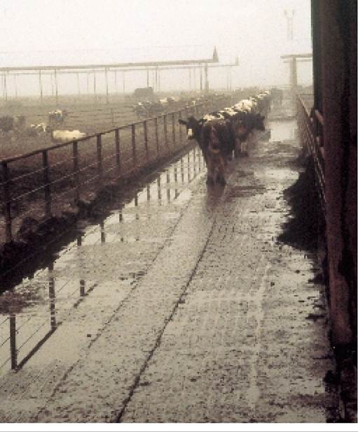
Plate 9.37. Proof that cows prefer to walk on a soft surface: they had the option of the whole width of concrete roadway, but preferred walking on the rubber belting in the centre. (Courtesy Karl Burgi.)
Management factors influencing lameness include:
• wet hooves, leading to soft horn
• poor foot surfaces
• rough handling
• inadequate or excessive hoof wear
• poor conformation
• routine foot trimming
• footbaths
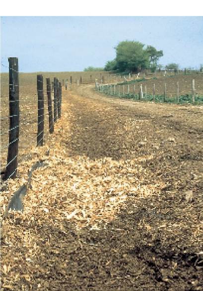
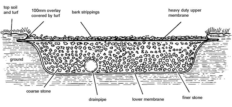
Figure 9.20. Construction of specific cow tracks provides a soft and comfortable walkway, helping to reduce lameness. (Courtesy John Hughes).
In the UK, if cows are allowed to amble out to a field at their own speed, they will usually choose to do so by walking on the soft earth of a grass verge, rather than on stones (Plate 9.38, right). They even place their feet in exactly the same spot each time, making holes in the ground. This preference for a softer surface has led to the development of specific cow tracks, as in Plate 9.38 (left) and Figure 9.20. The ground is excavated to 300 mm deep and 1-1.5 m wide and lined with a permeable geotextile membrane, a road construction membrane which prevents sinkage of the track. A drainage pipe runs along the base, surrounded by a large aggregate, perhaps having fine stone on the top. This is covered with a second special toughened membrane, which allows water to drain down through but will not allow mud to rise up through it. Finally a layer of bark strippings, sometimes
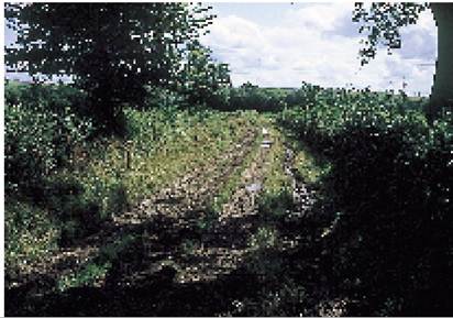
Plate 9.38. Specially constructed bark track will improve cow comfort and reduce lameness. (Courtesy Richard Cooke.) known as cundy peelings, 100 mm deep, is placed on top of the upper membrane to provide a comfortable walking surface for the cows. It must not be used by tractors and other vehicles.
Similar tracks may be constructed in gateways and around water troughs and in other areas where the ground gets badly poached. Plate 9.38 (left) shows a track running from the dairy down to fields half a mile away. Although the cows walked in almost single file along the track, they came up much more quickly than when they could only walk on the stony roadway. On very well drained land some farmers have constructed a simple track by scraping away the top soil and then unrolling a large round straw bale onto the underlying stone. Wet bales too badly soiled for straw yards can be used. The straw needs replacing approximately every two or three weeks, depending on the weather, but it makes a good track and is certainly cheaper.
The influence of floor surface on white line disease is interesting. It is commonly stated that cows become lame because of a specific type of stone or gravel in a track, particularly if sharp flints are present. However, beef cattle could almost certainly walk along the same track without the stones penetrating their feet - which suggests that it is the softening of the hoof and the weakening of the white line which are the critical factors and not the sharpness of the stones!
Rough handling Rough handling also has an effect. A survey of farms showed that cows which were forcibly rushed along farm tracks by a herdsman, dog or tractor had a far higher incidence of lameness than farms where the cows were allowed to walk along at their own speed. This was presumably because in the latter case they chose their own footing, thus minimising bruising to the sole and corium.
Hoof wear Both inadequate and excessive hoof wear can cause problems. Heifers reared and housed in totally bedded areas (straw, shavings or sand) do not get sufficient hoof wear. The toes become overgrown, the foot rotates backwards and the corium becomes damaged. The provision of a lightly abrasive concrete feeding area is essential. At the other extreme, cows or heifers (and especially fresh calvers) which are made to walk long distances on gravel or even concrete roads can wear their soles so thin that they are easily compressed by thumb pressure.
A similar ‘soft sole’ syndrome is seen in young bulls introduced to work in a dairy herd, particularly if the bulls are large and do not use the cubicles. The soles of their hind feet can wear down to the corium. Ideally bulls in cubicle systems should be rested in a straw yard, for example cubicles by day and a straw yard by night, or alternate weeks in cubicles and straw yards. On a daily basis bulls soon learn which is to be their period of lying down and compensate for the cubicles by lying down for long periods in the straw yards.
Conformation Conformation affects the incidence of lameness, which is therefore influenced by genetics and breeding. Bulls should be chosen to give a good depth of heel and a good upright angle of the front wall, as in Figure 9.7.
Foot trimming The final management factor which influences the degree of bruising of the corium is routine foot trimming - and this brings the discussion almost round in a full circle! If calving is a major stress period for the development of coriosis/laminitis, then feet need to be in optimum shape at calving in order to minimise this effect. This means trimming at drying off, especially removing overgrown toes and overgrowth of the sole, both of which could damage the fragile corium of the freshly calved cow.
The use of footbaths is discussed on page 318.
More on the topic SOLE ULCERS AND WHITE LINE DISEASE:
- Decubitus Ulcers
- Enforcing the Colour Line at Home and Abroad
- Reading between the Line
- No Single Line Across the Ocean
- Holding the Line Women, Ritual and the Protection of Rome[795]
- Democracy.. is the sole way of living which believes wholeheartedly in the process of experience as end and as means. John Dewey,from Creative Democracy
- Imperialism and ‘white rule' in southern Africa
- Geomyces Destructans - WHITE-NOSE SYNDROME IN HIBERNATING BATS
- INFECTIOUS BURSAL DISEASE (GUMBORO DISEASE)
- Imagine a situation where an African American family moves into an historically all white neighborhood.
- Norma White-Weithers, MS, DV
- White Blood Cell Disorders
- 21 White Bears, Whales and Walruses
- Jane A. Crowley and Kayla White-Water
- White Supremacist Terrorism in the United States