THE STRUCTURE OF THE FOOT
The bovine claw consists of three main components (Figure 9.1 and Plate 9.1). Moving inwards from the outer casing these are:
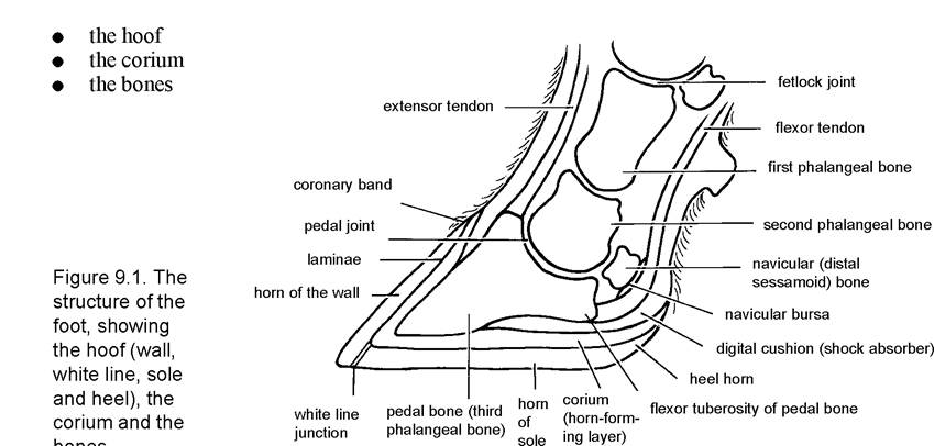
The Hoof
The hoof consists of four parts: the wall, the sole, the white line and the heel.
The wall is equivalent to the human finger-nail and is produced at the coronary band, that is the skin-horn junction at the top of the claw, shown in Figure 9.2. Once produced it flows down over the outside of the wall at approximately 5 mm per month. As the distance from the coronary band to the toe in the average cow is around 75 mm, this means that it can take 15 months (75 mm divided by 5 mm = 15) for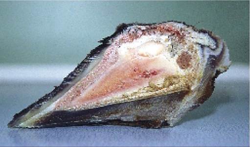
Plate 9.1. A cross-section of the hoof, showing horn of the wall, white line and sole, surrounding the corium and pedal bone.
new horn to come into wear at the toe. Towards the heel the average distance from the sole to the coronary band is only 30-40 mm (Figure 9.7), so horn produced at this point comes into wear more quickly. The coronary band is covered by the periople, which produces a smooth, waxy protective covering to the wall of the hoof. Damage to this leads to sandcracks.
The sole of the hoof is produced by the corium of the sole. The sole would be equivalent to a second ‘nail’ growing from the tip of a human finger. Where the wall joins the sole there is a ‘cemented’ junction known as the white line. This is clearly shown in Plate 9.1 and Figure 9.1. The white line runs from the heel along the outer (abaxial) wall of the hoof to the toe and then back for the first third of the inner (axial) wall (Figure 9.2). Because it is a cemented junction the white line is a point of weakness.
Whereas the wall and the sole consist of tubular horn (equivalent to concrete with steel reinforcing bars), the white line is more like cement. It has no tubular horn, it is less mature than the hornFigure 9.2. Diagram of right hind foot viewed from the bottom and the side, giving the nomenclature of its surfaces.
of the wall and it contains less keratin. All three factors make it much weaker and much more prone to injury.
The heel, or bulb of the hoof, consists of much softer horn and is produced by a continuation of the periople running from the coronary band. As it is a soft structure it expands and contracts during locomotion. This acts as both a shock absorber and a pump, thus allowing blood from the foot to be pumped back up the leg. Consequently if heifers (especially) spend too long standing still, the blood in the foot becomes ‘stale’ and this can result in poor horn formation. Alternatively excess trauma to the heel can sometimes produce a haematoma (blood blister), which causes lameness.
The Corium
The corium is the sensitive structure of the foot. A stone or nail penetrating the hoof causes pain and lameness only when the corium is compressed or penetrated. The corium is also a support tissue, carrying blood and nutrients to both the hoof and the pedal bone. When the corium is penetrated the foot may bleed. In addition to providing a nerve and blood supply, the corium is modified at various parts of the foot to provide three other important functions. These are:
• horn formation - by the papillae
• support for the hoof wall - by the laminae
• shock absorber and blood pump - by the digital cushion
Horn formation
Plate 9.2 shows the corium after the hoof has been removed. In the pale, cream-coloured area below the coronary band area and beneath, the corium is modified to form large numbers of finger-like projections
Plate 9.2. The hoof has been removed from this claw to expose the corium.
The lower reddish ‘fish gill' area is the laminae of the corium, the upper pale pink section the papillae (courtesy Dr. P. Ossent).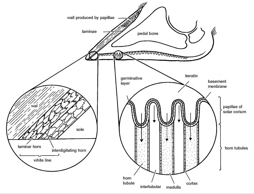
Figure 9.3. The structure of the hoof wall and the white line, showing details of horn formation.
known as the papillae. These are so small that they cannot be seen in Plate 9.2. The papillae extrude the tubular horn which eventually matures and hardens to form the hoof wall. Papillae are also found on the sole, where they extrude the horn of the sole. This is shown in detail in Figure 9.3. The corium also produces the cemented horn of the white line, but this time there are no papillae present, so there are no horn tubules and the horn is weaker.
Supportfor the wall
As the wall of the hoof provides weightbearing and protection for the foot, it has to be firmly attached to the underlying corium. However, at the same time it must be able to both move down over the foot and act as a shock absorber. The remarkable incorporation of these diverse functions is achieved using a series of interlocking leaves known as the laminae. These are clearly shown as the pink area in the lower part of the claw in Plate 9.2. Equivalent leaves are also present on the inside of the hoof wall (Plate 9.3) and these interdigitate with the laminae of the corium. A cow with laminitis has inflammation of the laminae. The increased blood flow, heat and swelling which this produces within the confined space of the hoof leads to intense pain and may distort the growth of the hoof. Although often referred to as laminitis, it is very rare that the laminae alone are affected. In most cows the whole of the corium will be inflamed, producing changes in the wall, sole and white line. The condition would therefore be best described as coriitis or coriosis.
The movement of the hoof wall down over the laminae has been compared to one piece of corrugated cardboard (the wall) moving down over a second stationary piece (the laminae of the corium).
This is shown diagrammatically in Figure 9.4.
Shock absorber and blood pump
Within the heel the corium is impregnated with fat, fibrous tissue and elastic material to form the digital cushion. The front portion of the digital cushion extends forward to run under the rear edge of the pedal bone, as shown in Figure 9.1. Because the heel horn is flexible, the digital cushion becomes compressed during weightbearing. When no longer weightbearing, the elastic tissue restores the cushion to its original shape. This regular expansion and contraction is important for both blood flow and shock absorption. If the corium becomes bruised or inflamed, due to excessive weightbearing or coriosis/laminitis respectively, the elastic and fat will be replaced by scar tissue. The function of the digital cushion (shock absorber and blood pump) will then be impaired.
The Bones
bones
There are really only two within the hoof, the pedal bone and the navicular bone. These are technically referred to as the third phalangeal bone and the distal sessamoid bone respectively. The pedal joint (distal interphalangeal joint), which is the junction between the second and third phalangeal bones, is also just within the hoof capsule, as can be seen in Plate 9.14 and Figure 9.1. Infection within this joint produces severe lameness.
F
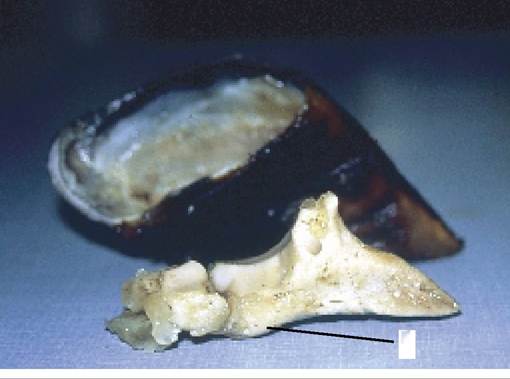
Plate 9.3. The pedal and navicular bones viewed from the inner aspect. Laminae can be seen on the inside of the hoof wall, and the flexor tuberosity F is clearly visible.
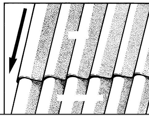
hoof wall
laminae of the corium
Figure 9.4. A diagrammatic representation of the laminae - one sheet of corrugated cardboard running over the other.
The navicular bone acts as a support structure, improving leverage by pushing the flexor tendon towards the heel as it runs down the leg and attaches to the base of the pedal bone.
The padded area between the flexor tendon and navicular bone is known as the navicular bursa (Figure 9.1).Although the pedal bone is weightbearing around its entire outer edge, its base is arch-shaped on its inner border. This is shown in Plate 9.3. The projection at the rear end of the pedal bone marked F in Plate 9.3 is the point of attachment for the flexor tendon. This projection is therefore called the flexor tuberosity. Compression of the corium between the flexor tuberosity of the pedal bone above and the hard horn of the sole beneath is an important part of the pathogenesis of sole ulcers and is referred to again on page 293. The corium feeds the bone as well as the hoof, so inflammation of the corium may also result in bone deformities.
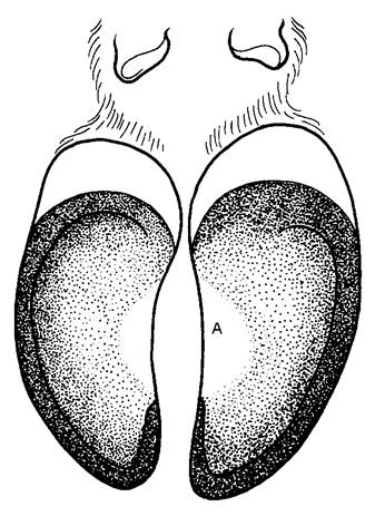
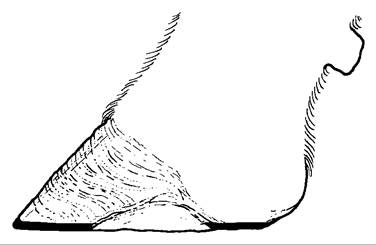
Figure 9.6. An axial (inner) view of the claw, showing weightbearing at the toe and heel, but not in the central sole area.
Figure 9.5. The correct weightbearing surfaces of the foot are the darker shaded areas. Note how the whole area of the toe is weightbearing.
More on the topic THE STRUCTURE OF THE FOOT:
- FOOT TRIMMING
- FOOT CONDITIONS CAUSING LAMENESS
- As we have seen, the legal structure of Roman marriage was fragile, in the sense that it was far from creating the family as a real partnership with respect to property; the wife appears, at times, almost as an intruder within a household structure still centered on the paterfamilias.
- OTHER CAUSES OF FOOT LAMENESS
- FOOT-AND-MOUTH DISEASE
- Immersion Injury (Trench Foot)
- CHAPTER 9 LAMENESS AND FOOT TRIMMING
- An Infeasible Assumption and Limited Information Hinder Veterinarian Workforce Planning Efforts for a Catastrophic Outbreak of Foot-and-Mouth Disease
- It was with “great wonderment” that Peter I learned of “the deed of the new Judas, Mazepa, who, after twenty-one years of loyalty to me and with one foot already in the grave, has turned traitor and betrayer of his own people.
- The structure of the legal standard.
- Biological structure of populations
- The general concept of structure
- The logical structure of econometrics