Removal
The softer wax balls can be curetted or flushed out of the ear canal with fluid under pressure. If there is an eardrum present, treatment with a ceruminolytic agent such as dioctyl sodium sulfosuccinate may loosen the concretion.
Warming the flush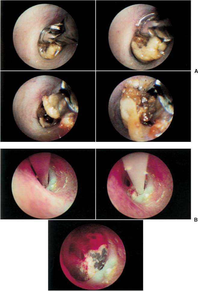
Figure 11-8
A, Removal of a wax ball from the ear canal from the cat in Figure 11-7. The mass was grasped by the endoscopic grasping forceps. Because the wax was quite soft and malleable, the endoscopic graspers were ineffective at removing the wax. B, The ear canal was treated with a ceru- minolytic agent and flushed with warmed dilute povidone-iodine solution. The soft wax was flushed from the ear and small pieces of remaining debris were carefully suctioned off the surface of the eardrum.
solution helps to soften the waxy debris, and sometimes careful suction with a large- bore suction catheter aids in their removal (Figure 11-8).
Removing a hardened concretion type of ceruminolith requires the use of a grasping forceps. The use of a video otoscope (Video Vetscope, MedRx, Inc., Seminole, Florida) facilitates this process because the veterinarian can see the ceruminolith clearly on the video monitor. The endoscopic grasping tool, which is inserted through the biopsy channel of the video otoscope, can be carefully placed to grasp and remove the mass (Figure 11-9).
After removal of these concretions, the tympanic membrane often looks abnormal. Small holes in the tympanum may be present because the epithelium is eroded along with the concretion as it is removed (Figure 11-10). These small holes heal rapidly (Figure 11-11).
If the eardrum was previously ruptured and a ceruminolith developed as a consequence, the healing tympanic epithelium may be in the middle of a ceruminolith.
When the concretion is removed, the eardrum is torn away, and a large direct communication to the tympanic bulla results. If detergent flush solutions were used for removal of the ceruminolith, copious saline flushes are used to remove these ototoxic substances (Figures 11-12 and 11-13).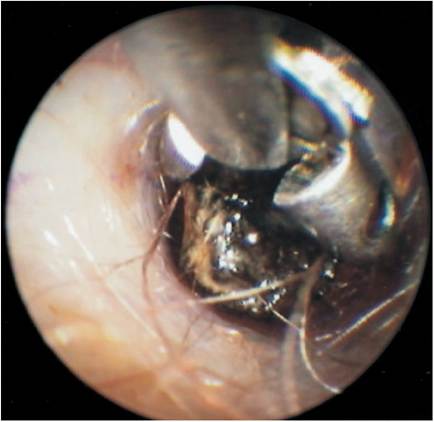
Figure 11-9
Endoscopic grasping forceps are used through the working channel of the Video Vetscope to extract the wax ball from the dog in Figure 11-5.
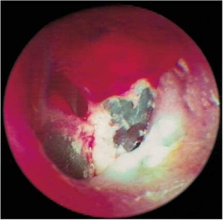
Figure 11-10
After removal of the wax plug from the cat in Figure 11-7, the eardrum was closely examined. A roughened surface and a small hole in the 5 o'clock position can be seen.
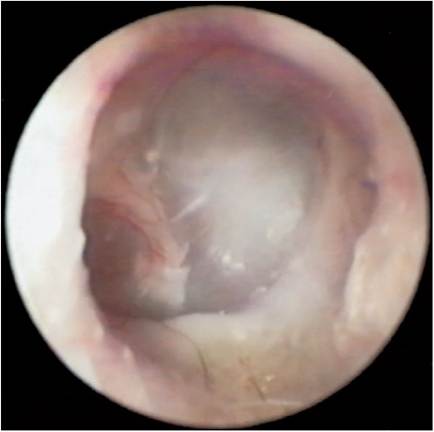
Figure 11-11
Close-up view of the eardrum from the cat in Figure 11-10 1 month after removal of the wax ball. The eardrum has healed and is almost normal in appearance. The cat was asymptomatic on this recheck visit.
Older animals may have large accumulations of waxy, cellular debris in the ear canals because the epithelial migration process is quite slow. Occasional ear flushes may be required to augment the physiologic cleaning process in these patients.
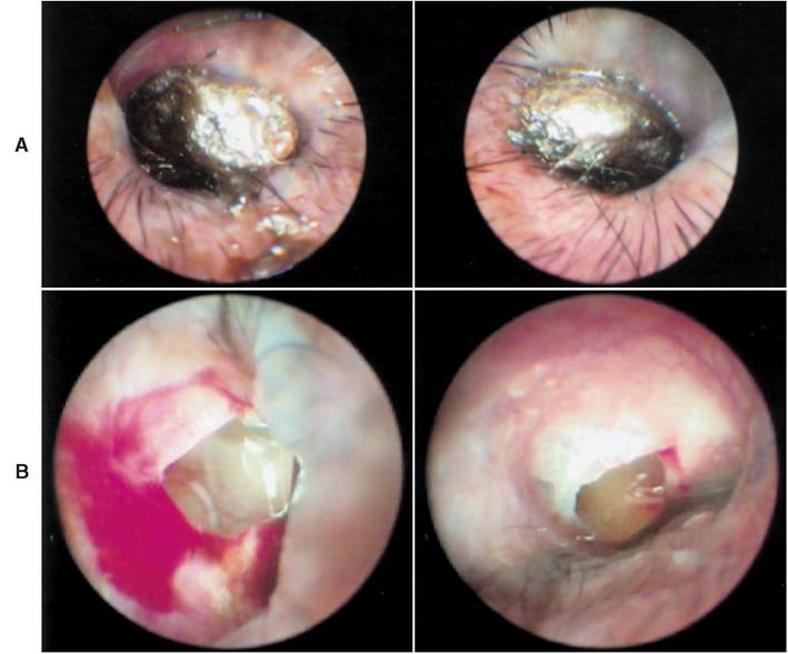
Figure 11-12
A, Bilateral Ceruminoliths were identified in a 7-year-old mixed-breed dog with diminished hearing. B, The Ceruminoliths were grasped with the endoscopic forceps and carefully removed. Both eardrums were ruptured, whiCh may have resulted beCause eaCh eardrum was involved in the matrix of the ceruminolith.
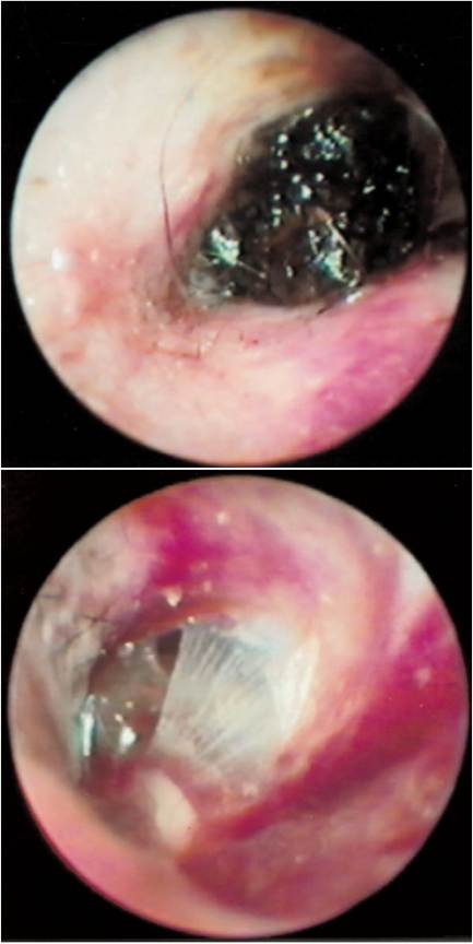
Figure 11-13
A, The patient, a 3-year-old German Shepherd, was examined because of a painful ear. The affected ear was not being carried in an erect position. Examination of the ear canal revealed a wax plug at the level of the eardrum. B, The wax plug was grasped with the endoscopic forceps and carefully extracted. After the ear canal was flushed and dried, a partial tear of the eardrum could be seen. Mucus and pus consistent with otitis media are present under the cerumen plug.
Suggested Readings
Michaels I, Soucek S: Development of the stratified squamous epithelium of the human tympanic membrane and external ear canal: the origin of auditory epithelial migration, Am JAnat 184:334344, 1989.
Smelt G, Stoney P, Weinberger J, et al: Sequelae of experimental tympanic and inferior wall perforations: the double meaning of epithelial migration, J Otolaryngol 20:171-176, 1991.
More on the topic Removal:
- 3 Fixtures and Buildings – Right of Removal
- 4 Removal of fruit trees, bushes, etc
- C REMOVAL OF SECURITY: THE CASES FOR POSSESSION
- Laser Polypectomy (Aural Tumor Removal)
- 6.57 Farming being capital intensive in nature, it is of central importance that the rights and liabilities of landlord and tenant as to the provision, repair, maintenance and removal of fixed equipment are clearly defined.
- Catchingup
- Plutarch and his effort to reject (and even ridicule) Epicurus and the Epicureans
- Control of BTB in Cameroon
- DISORDERS OF SPLEEN
- Answers to Analyzing Data 1.1 Questions
- Tombs and treatment of the dead
- The Hetmanate
- Conclusion
- Genital Warts
- The Earlier Republic (509-264 bc)
- Reasons for including resource dynamics