Tumors of the Ear Canal
Most tumors of the ceruminous glands of dogs are benign but in the cat are malignant. Ceruminous and sebaceous gland adenomas, adenocarcinomas, basal cell carcinomas, fibrosarcomas, chondrosarcomas, trichoepitheliomas, mast cell tumors, ceruminous gland hyperplasia, and inflammatory polyps have been identified in the ears of dogs and cats.
Nasopharangeal polyps have been identified in cats presented with otitis externa associated with ear masses. Tumors result in obstruction of the ear canal, with the most common ones being of ceruminous gland origin. Ceruminous gland tumors are most frequently diagnosed in older male cats; these tumors have a high malignancy rate.
Ceruminous gland adenomas (Figures 10-3 and 10-4) and carcinomas are the two most common tumors in the cat. The adenomas are smooth, nodular, or pedunculated masses with intact epithelium. If secondary infection is present, the epithelium may be ulcerated.
Histologically, adenomas are differentiated cystic or tubular growths with cuboidal eosinophilic epithelium (Figure 10-5). Cystic contents are colloidal, orange to eosinophilic secretions. The mass present in the ear canal may invade the parotid salivary gland. Inflammatory polyps in dogs and cats may be misdiagnosed clinically as neoplasia. Histopathologic diagnosis is necessary for differentiation of these masses.
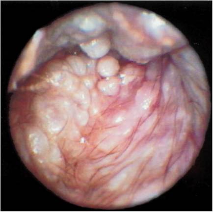
Figure 10-3
Small cerumen gland adenomas in the ear canal.
Figure 10-4
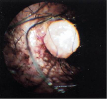
Large cerumen gland adenoma among cerumen gland hyperplasia.
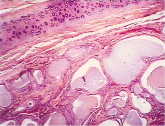
Figure 10-5
Histopathology of an ear canal, showing huge dilated cerumen glands and inflammation.
Clinical Signs.
Purulent, malodorous discharges are common clinical signs associated with any type of tumor in the ear canal. Secondary bacterial and Malassezia infections are common. Obstructions of the canal by these masses prevent drainage and result in the accumulation of debris and cerumen. The accumulation of this debris and cerumen causes irritation to the epithelium of the canal, resulting in hyperplasia and hypersecretion of the ceruminous glands. Sinusitis and dysphagia are often present in cats with nasopharyngeal polyps.Diagnosis and Tireatment. Otoscopic examination, preferably with the patient under general anesthesia, is necessary to determine the extent of these tumors. Although it is difficult to distinguish ceruminous gland hyperplasia from a neoplastic condition, it is important to determine the extent of the condition to plan for proper diagnostic procedures. For tumors of the ear, surgical intervention is the treatment of choice. Microscopic evaluation is necessary to make a definitive diagnosis of tumors in the ears of dogs and cats.
Histologic evidence of tumors (benign or malignant) warrants surgical intervention for tumor removal and proper ear drainage.
More on the topic Tumors of the Ear Canal:
- Careful examination of a clean, dry ear canal in a dog or cat with otitis externa may reveal many conditions that affect the ear canal.
- Lateral Ear Canal Resection and Ear Canal Ablation
- By definition, otitis externa represents a spectrum of inflammatory changes that occur to the external acoustic canal in response to any insult to the ear canal epithelium.
- Structure of the Ear Canal
- Flushing of the Ear Canal
- Vertical Ear Canal
- Examining the Ear Canal
- Histopatholog y of the External Ear Canal
- Horizontal Ear Canal
- The ear canal requires a clearance mechanism to remove the accumulation of dead cells, trapped foreign debris, and wax.
- Examination of the External Ear Canal
- BRAIN TUMORS
- SPINAL CORD TUMORS
- Tumors
- Tumors of the Bone
- HEPATIC TUMORS
- BONE MASSES AND TUMORS
- Testicular Cancer and Germ Cell Tumors