Structure of the Ear Canal
The external ear canal in the dog is 5 to 10 cm long and 4 to 5 mm wide (see Figure 1-5). The ear canal consists of an initial vertical part, which may extend an inch. The vertical canal runs ventrally and slightly rostrally before bending to a shorter horizontal canal that runs medially and forms the horizontal part of external ear canal.
Because the external ear is elastic, the ear canal can be straightened enough to permit otoscopic examination.The vertical part and most of the horizontal part of the canal are cartilaginous, but the deepest part is osseous. The ear canal is lined by skin containing sebaceous and ceruminous glands and hair follicles. The ceruminous glands are modified apocrine tubular sweat glands. The combined secretions of sebaceous and ceruminous glands constitute ear wax (cerumen). Cerumen (1) protects the external ear canal by immobilizing foreign objects and (2) keeps the tympanic membrane moist and pliable. The external ear canal is separated from the middle ear cavity by the semitransparent tympanic membrane.
Blood Supply to the External Ear
The external ear is generously vascularized by branches of the external carotid artery. A large caudal auricular artery arises from the external carotid artery at the base of the annular cartilage, medial to the parotid salivary gland and deep to the caudal
CLINICAL NOTE
Violent shaking of the head by the animal may contribute to fracture of the delicate auricular cartilage, resulting in severe hemorrhage within the cartilage. The blood clot (aural hematoma) may often fill the entire concave surface of the ear, requiring surgical removal of the clot.
auricular muscles. This artery gives off the lateral, intermediate, and medial auricular arteries, which pass along the convex surface of the pinna, wrapping around the helicene margins as well as penetrating the scapha and supplying the skin covering the cavum conchae.
In addition to providing nourishment to the tissues of the external ear, the vascular supply to the pinna may also play a minor thermoregulatory role. Venous drainage occurs via the caudal auricular and superficial temporal veins into the maxillary vein.Nerves of the External Ear
Sensory innervation of the pinna and external ear canal is provided by four nerves: the trigeminal, facial, vagus, and second cervical.
The auriculotemporal branch of the trigeminal nerve provides sensory innervation to the skin lining the horizontal part of the ear canal and to the tympanic membrane itself. This nerve also provides sensory innervation to the rostral margin of the pinna and the concave surface of the pinna close to the rostral margin and the skin over the tragus.
The facial nerve is related to the ventral surface of the annular cartilage, close to the osseous external acoustic meatus. The facial nerve provides substantial sensory innervation to the concave surface of the scapha and part of the cavum conchae via the rostral, middle, and caudal internal auricular branches (Figure 1-6). Most of the vertical along with part of the horizontal ear canal lining is supplied by the lateral internal auricular branch of the facial nerve, which may contain predominantly vagal fibers.
Communication between the facial nerve and the vagal nerve takes place as the facial nerve exits the stylomastoid foramen (see Figure 1-11). It is believed that these vagal branches are given off as the lateral internal auricular branch to the skin of the external ear canal. Reflex gastric vomiting may be triggered if the sensory endings of the vagus nerve are stimulated by mild ear canal irritation.
The convex surface of the pinna is provided with sensory innervation mainly by the second cervical nerve. All of the muscles of the external ear are innervated by the facial nerve.
Tympanic Membrane
The tympanic membrane (eardrum) is a thin, slightly opaque, membranous partition that separates the external ear from the middle ear.
The tympanic membrane is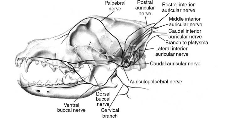
Figure 1-6
Schematic drawing of sensory branches of the facial nerve that supply the external ear. Other important motor branches of the facial nerve are also shown.
located at a 45-degree angle in relation to the central axis of the horizontal part of the external ear canal (Figure 1-7; see also Figure 1-5). It is thin in the center and thicker near the periphery. The small upper portion is the pars flaccida, and the larger lower part is the pars tensa (Figure 1-8). With the exception of the pars flaccida, the membrane is tense, being firmly attached to the surrounding bone by a fibrocartilaginous ring, the annulus fibrocartilaginous. This ring is attached to the osseous ring of the external acoustic meatus by fibrous tissue.
The pars flaccida is a loose, opaque, pink triangular region forming the upper quadrant of the eardrum containing small branching blood vessels. Owing to its flaccid nature and rich blood supply, the pars flaccida heals rapidly if injured.
The pars tensa is thin, tough, and glistening, usually pearl-gray and translucent, although it may have opaque, radiating strands. The pars tensa, once broken, heals slowly. The external aspect of the tympanic membrane is concave because of traction on the medial surface by the manubrium of the malleus. The outline of the manubrium of the malleus is usually visible through the tympanic membrane as the stria mallearis (see Figure 1-8).
Opposite to the distal end of the manubrium, the depressed point on the external surface of the tympanic membrane is called the umbo membrane tympani.
Histologically, the tympanic membrane is made up of four layers: an external epidermal layer, an inner mucous layer, and two layers of intervening fibrous tissue. The epidermal layer is made up of a thin hairless skin consisting of a flat basal layer without any ridges, and a superficial layer only a few cells thick.
This stratified squamous epithelium is continuous with the epithelial lining of the external ear canal. The thin dermis contains fibroblasts and a fine vascular supply. The thicker middle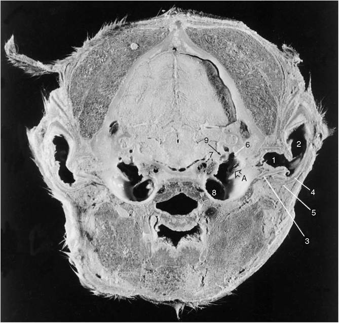
Figure 1-7
Transverse section through the head of the dog at the level of the tympanic bulla. A, Osseous external acoustic meatus covered by tympanic membrane. Note that the tympanic membrane is placed at an approximately 45-degree angle in relation to the central axis of the horizontal part of the ear canal. 1, Horizontal part of the ear canal; 2, vertical part of the ear canal; 3, annular cartilage; 4, auricular cartilage; 5, parotid salivary gland; 6, epitympanic recess; 7, carotid canal accommodating the internal carotid artery, postganglionic sympathetic nerves, and the ventral petrosal venous sinus (the caudal continuation of cavernous venous sinus), which drains into the internal jugular and vertebral veins; 8, cavity of the middle ear; 9, osseous labyrinth accommodating the internal ear.
fibrous layer consists of an outer layer of fibers that radiate toward the annulus fibrocartilaginous from the center of the eardrum. A layer of inner fibers are circular and are found closer to the annulus. The manubrium of the malleus is buried in the fibrous layer intercalated between the two epithelial surfaces. The arrangement of fibers in the middle layer optimizes the vibratory faces. The arrangement of fibers in the middle layer optimizes the vibratory response of the tympanic membrane to incoming sound waves. The inner layer of the tympanic membrane is composed of
-AfcT ∙
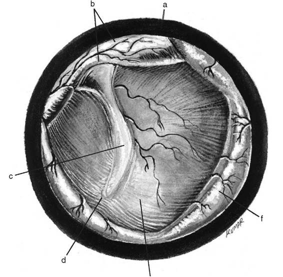
Figure 1-8
Tympanic membrane anatomy in the dog as observed through an otoscope. a, Rim of the otoscopic tube; b, flaccid part of the tympanic membrane; c, manubrium of malleus shining through the tympanic membrane; d, umbo of tympanic membrane; e, pars tensa of the tympanic membrane surrounding the manubrium; f, skin of the external acoustic meatus raised by the otoscopic tube. (Modified from DeLahunta A, Habel RE: Applied veterinary anatomy, Philadelphia, 1986, WB Saunders.)
a single layer of respiratory epithelium. The epithelium lining the inner surface of the tympanic membrane starts as columnar at the periphery, gradually becoming cuboidal and finally squamous at the center. This layer is continuous with the mucous membrane type of respiratory epithelium covering the middle ear cavity, auditory tube, and nasal cavity. The underlying lamina propria is thin, with a fine vascular supply.
More on the topic Structure of the Ear Canal:
- Careful examination of a clean, dry ear canal in a dog or cat with otitis externa may reveal many conditions that affect the ear canal.
- Lateral Ear Canal Resection and Ear Canal Ablation
- By definition, otitis externa represents a spectrum of inflammatory changes that occur to the external acoustic canal in response to any insult to the ear canal epithelium.
- Flushing of the Ear Canal
- Vertical Ear Canal
- Examining the Ear Canal
- Histopatholog y of the External Ear Canal
- Horizontal Ear Canal
- Tumors of the Ear Canal
- The ear canal requires a clearance mechanism to remove the accumulation of dead cells, trapped foreign debris, and wax.
- Examination of the External Ear Canal
- Structure and Functioning of Ear
- Structure of the External Ear
- As we have seen, the legal structure of Roman marriage was fragile, in the sense that it was far from creating the family as a real partnership with respect to property; the wife appears, at times, almost as an intruder within a household structure still centered on the paterfamilias.